Article
Anal fissure
- Steven Schlichtemeier, Alexander Engel
- Aust Prescr 2016;39:14-7
- 1 February 2016
- DOI: 10.18773/austprescr.2016.007
An anal fissure is a common, mostly benign, condition that can be acute or chronic. The diagnosis is usually made on history and physical examination, but further investigations are sometimes necessary.
Primary fissures are usually benign and located in the posterior or anterior position. Secondary fissures are lateral or multiple and often indicate a more serious underlying pathology.
The management of primary anal fissures is generally non-operative and includes increased dietary fibre, sitz baths, topical ointments and botulinum toxin injections. If these treatments are ineffective the patient will need a surgical referral.
Secondary anal fissures require further investigation. Multidisciplinary management is preferable and is essential in the case of malignancy.
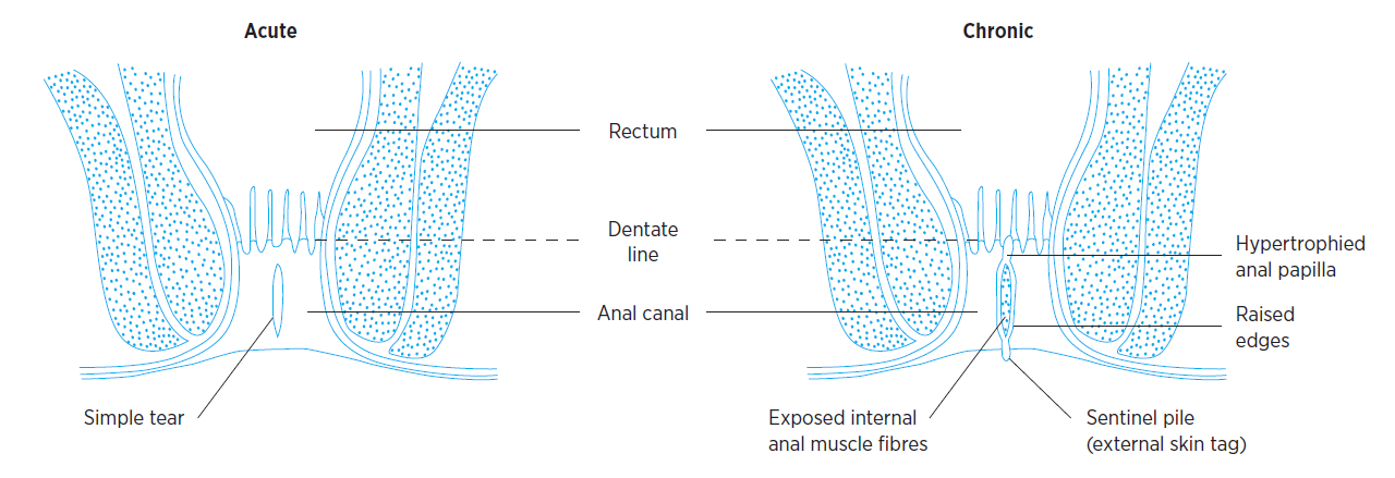
An anal fissure is a longitudinal tear or defect in the skin of the anal canal distal to the dentate line (Fig. 1). The classification of anal fissures is based on causative factors.
Primary fissures are typically benign and are likely to be related to local trauma such as hard stools, prolonged diarrhoea, vaginal delivery, repetitive injury or penetration. Secondary fissures are found in patients with previous anal surgical procedures, inflammatory bowel disease (e.g. Crohn’s disease), granulomatous diseases (e.g. tuberculosis, sarcoidosis), infections (e.g. HIV/AIDS, syphilis) or malignancy.1
An acute anal fissure commonly heals with 4–8 weeks of conservative therapy. If this therapy fails and the fissure becomes chronic, surgery is usually required.2-4
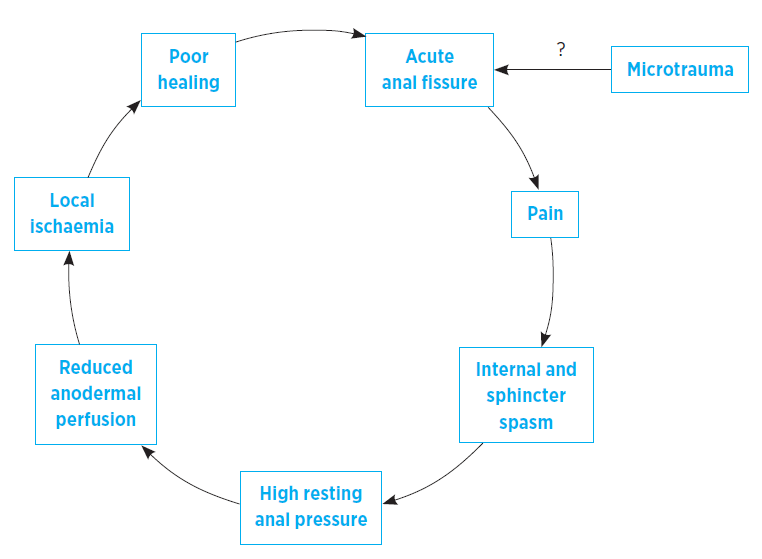
The pathophysiology of anal fissures is not entirely clear. It is probable that an acute injury leads to local pain and spasm of the internal anal sphincter. This spasm and the resulting high resting anal sphincter pressure5 leads to reduced blood flow and ischaemia,6,7 and poor healing. Unless this cycle is broken the fissure will persist (Fig. 2).
In approximately 90% of patients the anal fissure is located in the posterior midline. It is hypothesised that this predilection for the posterior midline may occur because this portion of the anal canal is poorly perfused.7,8 Anterior anal fissures affect approximately 10% of patients and may have a different pathophysiology. They are associated with younger, mostly female, patients often with injury to or dysfunction of the external anal sphincter. In less than 1% of patients the fissures are lateral or multiple.2 Irrespective of these differences posterior and anterior anal fissures are thought to be of primary aetiology, whereas lateral or multiple fissures are more likely to be secondary in nature.2
A small study of completely excised anal fissures found no underlying microscopic features of inflammation in most of the patients. Further, these fissures or defects showed little in the way of ulcer characteristics and appeared to be more consistent with unstable anodermal scar tissue.9 Additional research is needed to understand the temporal relationship between poor perfusion and lack of inflammation, as well as to identify the best terminology to describe these lesions.
History and physical examination will allow the diagnosis of an anal fissure without further investigations in most patients. The clinical features are severe tearing pain with the passage of faeces often with a small amount of bright red blood on the stool or toilet paper. The ideal way of examining is to have the patient lie comfortably in a lateral position and then gently part the buttocks to look first at the posterior midline.
An acute anal fissure appears as a fresh laceration, while a chronic anal fissure has raised edges exposing the internal anal sphincter muscle fibres underneath. Chronic anal fissures are also often accompanied by an external skin tag (sentinel pile) at the distal end of the fissure and a hypertrophied anal papilla at the proximal end (difficult to see on physical examination) (Fig. 1).
A digital rectal examination is usually not needed to make the diagnosis and is contraindicated in many cases given the associated pain. However, examination under anaesthesia with anoscopy, endoscopy, biopsy and imaging (i.e. CT scan, MRI or endoanal ultrasound) may all be required if:
The differential diagnosis of a primary anal fissure is limited but includes a haemorrhoid, anal fistula or solitary rectal ulcer. These conditions can be excluded by careful clinical assessment.
Secondary anal fissures may have characteristic features in the patient’s history such as risk factors for anal cancer, or medical conditions such as Crohn’s disease, tuberculosis, sarcoidosis, HIV/AIDS and syphilis. These fissures often lie laterally or are multiple in number. Further investigations must be performed as the underlying cause will determine subsequent management.
There are no clear guidelines on anal fissure management. The goals of management are to break the cycle of anal sphincter spasm allowing improved blood flow to the fissured area so that healing can occur. Almost 50% of patients with acute anal fissures will heal with conservative measures alone involving only increased fibre intake (e.g. psyllium) and warm bathing of the perineum (sitz baths).4,10 It is hypothesised that warm baths lead to relaxation of the internal anal sphincter via a somatoanal reflex.11
First-line therapy often includes the conservative measures plus a topical drug. The preparations used in clinical practice contain glyceryl trinitrate or a calcium channel blocker.
A recent Cochrane review reported that topical glyceryl trinitrate is better than placebo in healing anal fissures (healing rates 49% vs 36%). However, late recurrence occurred in around 50% of those initially cured. It also reported that calcium channel blockers (pooling results from studies using topical or oral preparations) had comparable efficacy to topical glyceryl trinitrate.12 One study from this review reported that topical diltiazem has superior healing rates to oral diltiazem (65% vs 38%).13 While topical diltiazem is the most predominantly studied and clinically used calcium channel blocker, topical nifedipine has also shown some encouraging results.14
The typical dosing of either 0.2% nitroglycerin ointment or 2% diltiazem cream is twice daily for 6–8 weeks.4 Topical glyceryl trinitrate is believed to work through its metabolites. It breaks the cycle of spasm by relaxing the internal anal sphincter and reducing resting anal pressure. Topical calcium channel blockers also relax the internal anal sphincter by blocking the influx of calcium into smooth muscle cells.
The main limitation to using topical glyceryl trinitrate is headaches and lightheadedness. This results in up to 20–30% of patients ceasing therapy prematurely.2,12 Headaches also occur in a similar proportion of patients using topical calcium channel blockers, however they occur less frequently so may be more tolerable.3
Patients using topical glyceryl trinitrate should not take sildenafil, tadalafil or vardenafil due to the risk of hypotension. For patients with angina or heart failure taking nitrates, topical glyceryl trinitrate may cause nitrate tolerance if used during the nitrate-free interval.15
Other topical medications commonly used in clinical practice are lignocaine and hydrocortisone. However, they have inferior healing rates to bran plus warm sitz baths.16 There are also several other topical medications under investigation including bethanechol, indoramine, minoxidil, clove oil and sildenifil, but current evidence does not support their use.4 Current evidence also does not support the use of oral rather than topical calcium channel blockers in the management of anal fissures.12
The reported healing rates of anal fissure following botulinum toxin injection are 60–80% (superior to placebo). Although recurrence can occur in up to 42% of patients, repeated injection has similar healing rates. Common adverse effects include temporary incontinence of flatus (in up to 18%) and stool (in up to 5%).4 The available evidence suggests that these injections probably have at least similar efficacy (certainly not worse) to both topical glyceryl trinitrate and calcium channel blockers.12,17
In clinical practice, given the invasiveness of these injections and the adverse-effect profile, some clinicians use botulinum toxin as second-line therapy, particularly in high-risk patients (young multiparous females with reduced sphincter mass), before referring them for a surgical opinion. However, other than the common adverse effects, the main disadvantage with botulinum toxin is that there is no consensus on the number of units to inject or the preferred location for these injections. This makes it difficult to interpret the variable healing rates published in the literature.
Surgery is considered for patients not responding to conservative measures. Although the timing of surgery is individual and variable, the literature often suggests between 4 and 12 weeks (6–8 weeks may be the ideal timing) after starting conservative treatment given the recommended duration of some of the topical dosing regimens.
The gold standard surgical operation for anal fissure is lateral internal sphincterotomy. This procedure commonly involves division of the internal anal sphincter from its distal end to either the proximal end of the fissure or the dentate line (whichever comes first). Lateral internal sphincterotomy has an excellent healing rate of approximately 95%. Common complications include recurrence in up to 6% and incontinence of flatus or stool (usually transient) in up to 17% of patients.12
When comparing lateral internal sphincterotomy to the historical four-finger anal stretch, lateral internal sphincterotomy is superior both in terms of recurrence and minor incontinence. However, a more standardised approach using pneumatic balloon dilation has shown healing rates of 83%, approaching those of lateral internal sphincterotomy, but with a lower incidence of long-term incontinence.1
When comparing lateral internal sphincterotomy to topical glyceryl trinitrate, calcium channel blockers and botulinum toxin injection, lateral internal sphincterotomy is clearly superior in terms of healing rates. However, it has more complications in some but not all studies.18-20
In recent years there has been growing interest in spinchter-sparring surgical techniques, predominantly that of fissurectomy either alone or in combination with other techniques (e.g. botulinum toxin injection or advancement flap). One observational study with good long-term follow-up reported that simple fissurectomy had a healing rate of 88%, a recurrence rate of 11.6% and an incontinence rate of 2.3%.21 Although not as successful or durable as lateral internal sphincterotomy, some would argue this to be more than a fair trade-off given the preservation of the sphincter complex and hence much lower incontinence rate.
A high index of suspicion is warranted for fissures in lateral or multiple locations and those not healing despite conservative therapies. Once investigated and diagnosed, management of secondary fissures will involve an extensive multidisciplinary approach involving gastroenterologists, infectious disease specialists, oncologists, pathologists and colorectal surgeons. Although surgery may ultimately benefit some patients with inflammatory bowel disease or HIV/AIDS, it may be contraindicated if there is malignancy.
The management of primary anal fissures usually follows a step-wise approach with first-line medical therapy for up to 6–8 weeks. Botulinum toxin injections may be reserved for second-line therapy although they may be used in combination with the conservative therapies. Patients not responding to these measures should be referred for surgery. In the case of a suspected secondary anal fissure, surgical therapies should be postponed or avoided depending on the results of further investigations and multidisciplinary management.
Colorectal fellow, Royal North Shore Hospital
Clinical academic, Royal North Shore Hospital
Associate professor, Surgery, Northern Clinical School, University of Sydney