Article
The assessment of severe cutaneous adverse drug reactions
- Ana M Copaescu, Jason A Trubiano
- Aust Prescr 2022;45:43-8
- 1 April 2022
- DOI: 10.18773/austprescr.2022.010
Severe cutaneous adverse drug reactions include Stevens-Johnson syndrome, toxic epidermal necrolysis and acute generalised exanthematous pustulosis. These eruptions are a type of delayed hypersensitivity reaction and can be life-threatening.
The assessment of a severe cutaneous drug reaction requires a detailed clinical history and examination to identify the culprit drug and evaluate the allergy. Allopurinol, antibiotics and anticonvulsants are often implicated.
Patch testing and delayed intradermal testing can assist in determining if the reaction was allergic, however there is limited evidence about the sensitivity and specificity of skin testing in severe cutaneous adverse drug reactions. If the testing is non-conclusive or negative, it is recommended to avoid the suspected culprit drug and any structurally similar drug in future.
Any decision to reintroduce a drug should be made after considering the harm–benefit ratio. Caution is also needed if considering a possibly cross-reactive drug in a patient with a history of severe cutaneous adverse drug reactions.
Skin eruptions can occur during drug treatment. They have a variety of causes including drug hypersensitivity. In allergic drug reactions, the immune system is triggered by a drug. These allergic reactions are unpredictable and not necessarily dependent on the dose.1 In Australian primary care, 10% of the encounters are for an adverse drug event among which 11% are considered related to an allergic reaction.2
The immediate type of drug hypersensitivity reaction occurs soon after exposure to the drug. It is thought to be mediated by immunoglobulin E. In contrast, severe cutaneous adverse reactions are due to delayed drug hypersensitivity and are presumed to be T-cell mediated.3 These immune-mediated reactions cause severe damage to the skin (Fig.) and internal organs, and are associated with significant acute and long-term morbidity and mortality. Allopurinol, antibiotics and anticonvulsants are often implicated.4
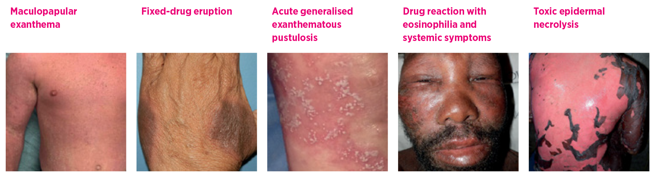
Adapted, with permission from Elsevier, from reference 13
Table 1 lists the severe cutaneous adverse reactions to drugs.3 These include acute generalised exanthematous pustulosis, drug reaction with eosinophilia and systemic symptoms (DRESS, also known as drug-induced hypersensitivity syndrome) and Stevens-Johnson syndrome. The Stevens-Johnson syndrome and toxic epidermal necrolysis are thought to be variants of the same condition. Mortality rates can reach 30–50%.5 The distinction between Stevens-Johnson syndrome and toxic epidermal necrolysis is determined by the affected body surface area:
Table 1 - Diagnostic tests and scoring algorithms for assessing delayed drug hypersensitivity reactions
|
Acute generalised exanthematous pustulosis |
Drug reaction with eosinophilia and systemic symptoms |
Stevens-Johnson syndrome/ toxic epidermal necrolysis |
|
|
Clinical manifestations |
Non-follicular sterile pustular rash over widespread erythema, fever and laboratory abnormalities* |
Erythematous urticaria-like or violaceous skin eruption, facial and extremity oedema, lymphadenopathy, fever, laboratory abnormalities* and internal organ involvement |
Skin necrosis, skin detachment and blistering of the mucous membranes accompanied by serious systemic manifestations |
|
Commonly implicated drugs3 |
Antibiotics (penicillins, cephalosporins) Antimycotics Other (diltiazem, antifungals, analgesics) |
Anticonvulsants Antibiotics (sulfonamides, vancomycin, minocycline) Allopurinol |
Allopurinol Anticonvulsants Antibacterial sulfonamides Nevirapine NSAIDs Antituberculosis drugs |
|
Scoring algorithms |
|||
|
Disease likelihood |
AGEP validation score |
RegiSCAR score |
n/a |
|
Drug causality |
Naranjo score |
Naranjo score |
ALDEN score Naranjo score |
|
Mortality prediction |
SCORTEN |
||
|
Diagnostic tests |
|||
|
Patch testing |
Indicated |
Indicated |
Indicated |
|
Delayed intradermal testing |
Indicated |
Indicated |
NOT indicated |
|
Oral challenge |
NOT indicated |
NOT indicated |
NOT indicated |
* Laboratory abnormalities refer to biochemical abnormalities such as increased concentrations of creatinine and liver enzymes (aspartate aminotransferase, alanine aminotransferase) or haematological abnormalities such as eosinophilia and neutrophilia.
NSAIDs non-steroidal anti-inflammatory drugs
AGEP acute generalised exanthematous pustulosis
RegiSCAR European registry of severe cutaneous adverse reactions
n/a not applicable
Naranjo adverse drug reaction probability scale
ALDEN algorithm of drug causality for epidermal necrolysis
SCORTEN score of toxic epidermal necrosis
Several clinical manifestations should raise the suspicion of a severe cutaneous adverse reaction. These include dark-purple skin infiltration, facial swelling, skin peeling and blistering, mucosal involvement, adenopathy, fever and haematological and biochemical laboratory abnormalities. A presence of any of these should warrant urgent hospital referral.
An adverse event that involves a drug should be reported to the Australian Therapeutic Goods Administration.
The most common benign cutaneous reaction to drugs is the maculopapular exanthema or morbilliform drug eruption. This is characterised by maculopapular red skin lesions that can become widespread and confluent. There may be pruritus and mild eosinophilia.3
The fixed-drug eruption is a reaction characterised by well-defined, red–dark, burning or itchy lesions. These lesions may reappear in the same areas on re-exposure to the drug.5 In a generalised bullous fixed-drug eruption there are sharply defined bullae at the same site following recurrent administration of the offending drug.6
Another drug eruption is symmetrical drug-related intertriginous and flexural exanthema. This is a well- demarcated macular eruption involving the flexural or intertriginous folds, and inguinal and perigenital as well as the gluteal and perianal areas.7
While technically not severe cutaneous adverse reactions, drug-induced liver injury and acute interstitial nephritis are examples of possibly severe single-organ diseases that can have pruritic skin eruptions.
Another multisystem disease related to drug exposure is the abacavir hypersensitivity syndrome. This is characterised by skin eruption, fever and gastrointestinal symptoms usually in the first weeks of therapy.8,9
Some tools have been developed to help establish the likelihood of a particular reaction (Table 1). Examples include tools for the diagnosis of acute generalised exanthematous pustulosis,10 DRESS11 and Stevens-Johnson syndrome or toxic epidermal necrolysis.12
In some cases of atypical skin lesions, a skin biopsy could be performed. However, there are no definitive histological criteria for the diagnosis of drug-induced eruptions and a skin biopsy might not exclude alternative causes for the eruption. Biopsy is supportive but not definitive.
As patients are often taking numerous drugs, evaluating drug causality in severe cutaneous adverse reactions can be challenging.4,5,13-15 The initial assessment includes constructing a drug timeline from the patient’s history and a detailed review of any drugs started in the 6–8 weeks before the reaction occurred. Generally, drugs started eight weeks before the skin eruption are not implicated. Common offenders include:
Some of the severe cutaneous adverse reactions present with constitutional symptoms, so one must keep in mind that some of the drugs given to treat these early symptoms might be incorrectly considered to have caused the eruption. Using validated drug causality tools (Table 1) such as the Naranjo score16 can help to minimise this error. This simple and widely used scale is reserved for the evaluation of adverse drug reactions.17 A Naranjo score of 4–5 is likely to indicate drug causality.
These tools help to categorise the most likely causal drug, considering the type of drug, the timing and possible alternative causes.18 If the repeated administration of a suspected drug has caused no symptoms, that drug may be excluded as a possible offender. Similarly, recurrent symptoms that present following the administration of the same drug would increase the likelihood that it caused the reaction. If similar signs and symptoms have occurred in the absence of any medicine, a non-drug-related condition should be considered in the differential diagnosis.
Some specialised centres are developing new laboratory tools, which examine cytokine production from isolated patient T cells. These aim to help evaluate drug causality, however their use is currently reserved for research purposes.19-21
Following complete resolution of the acute reaction, various investigations are available in specialised centres. These are generally performed at least six weeks after the complete resolution of the acute disease or after stopping immunosuppressive treatment.19,22
Patch testing involves applying a diluted sterile concentration of the implicated drug in a soluble medium under occlusion on the patient’s skin, to see if the initial reaction is reproduced in that small testing area. This is a quick and safe investigational method and is clinically relevant if the result is conclusive. A negative patch test does not exclude the drug as a possible cause.23 For severe delayed immune-mediated reactions, such as Stevens-Johnson syndrome or toxic epidermal necrolysis, patch testing should be delayed for six months after the resolution of the skin reaction.24
Intradermal testing with delayed reading (48–72 hours) can be done with various non-irritating concentrations of sterile commercially manufactured preparations.22 These are injected into the forearm. Like patch testing, intradermal testing should be performed at least four to six weeks after an acute reaction. The ability of delayed intradermal testing to detect true cases of allergy varies. Its sensitivity for antimicrobials ranges from 6.6–36.3% for cases of maculopapular exanthema to 64–100% for DRESS.25 In our Australian experience, intradermal testing has identified the causative drug in 46–56%, particularly for cases of severe maculopapular exanthema and DRESS.19,26
Safety considerations and the low described sensitivity and specificity of intradermal testing and patch testing limit their use in the management of severe cutaneous adverse reactions.22,27,28 Considering the limited number of diagnostic tools for the assessment of these very severe conditions, skin testing is still considered an essential clinical tool for providing guidance to clinicians. Conclusive results on skin testing will help to identify alternative drugs for patients who have multiple allergies.
The gold standard for drug allergy assessment is drug rechallenge. Depending on the availability of the implicated drug, a rechallenge can be performed with oral, intravenous or intramuscular doses. However, a rechallenge is not without risk and there are often other drug alternatives. The majority of local and international guidelines advise against a drug rechallenge in patients who have had severe cutaneous adverse reactions.
Cross-reactivity is when an individual, previously exposed and allergic to a drug, is exposed to a structurally similar drug, and the immune system recognises the shared chemical structure resulting in an allergic reaction.
The majority of the data on cross-reactivity come from immediate rather than delayed hypersensitivity.
When a patient is allergic to a drug and the alternatives are limited or associated with adverse drug reactions, allergy investigations are suggested. Skin testing can be performed with the implicated and cross-reactive drugs. If skin testing is positive in the setting of a severe systemic reaction, the tested and structurally similar drugs must be avoided. A similar approach is recommended in the setting of a non-conclusive test and there must always be a consideration of the harm–benefit ratio.
The most common example of cross-reactivity is among the penicillin family of antibiotics. However, the label of penicillin allergy may be incorrect.29 According to studies on delayed hypersensitivity reactions, among a cohort of patients with positive patch testing or intradermal testing to at least one penicillin reagent, none of the patients reacted to carbapenems.30 Following specialist consultation, carbapenems could be considered for a patient with a history of a severe cutaneous adverse reaction to penicillin. If the initial reaction was to an aminopenicillin, the recommendation is to avoid all aminocephalosporins sharing a similar side chain, such as cefalexin and cefaclor.25 Following an assessment of the allergy, these patients could be able to tolerate other cephalosporins.31,32 Cefazolin has no common side chains with other molecules and is regularly tolerated by patients with a penicillin or cephalosporin allergy – however, specific data regarding severe cutaneous adverse reactions are lacking.
In patients labelled allergic to sulphonamides such as the trimethoprim and sulfamethoxazole combination, studies have reported that there is no cross-reactivity between antibacterial (e.g. sulfasalazine and sulfamethoxazole) and non-antibacterial sulphonamides (e.g. acetazolamide, furosemide (frusemide), celecoxib, thiazide diuretics, sumatriptan, sotalol, probenacid).25 This lack of cross-reactivity has also been reported for cases of severe cutaneous adverse reactions.33 However, there seems to be cross-reactivity between dapsone and trimethoprim/ sulfamethoxazole and caution is advised.34,35
Cross-reactivity has also been reported among the drugs belonging to the families of macrolides, tetracyclines, aminoglycosides, quinolones, glycopeptides and nitroimidazoles.25
Allopurinol can cause a maculopapular drug eruption and severe cutaneous adverse reactions such as DRESS, Stevens-Johnson syndrome and toxic epidermal necrolysis with an overall incidence of 2%.36 The median time of onset is three weeks, but some reactions have been reported several years after starting treatment.37 In patients who have an indication for urate-lowering treatment (e.g. gout, hyperuricaemia and tumour lysis syndrome) and who have had a severe reaction to allopurinol, alternative drugs should be considered. Some studies have described desensitisation regimens and the harms and benefits of these should be discussed with an allergy specialist.38
Patients who have reacted to aromatic antiepileptic drugs, such as carbamazepine, oxcarbazepine, phenytoin, phenobarbital, lamotrigine, felbamate and zonisamide, should avoid all the drugs of this specific family. However, there is evidence that these patients will tolerate valproic acid and structurally distinctive anticonvulsants, such as benzodiazepines (e.g. clobazam, clonazepam) and gabapentin.39
There are specific genetic associations between human leukocyte antigen (HLA) alleles and severe cutaneous adverse reactions. These discoveries have increased the understanding of the immune mechanisms of delayed hypersensitivity reactions and enabled the development of screening guidelines and specific programs (Table 2).13,40,41
Table 2 - Genetic screening in delayed immune-mediated adverse drug reactions
|
Drug |
Severe cutaneous adverse reactions |
Human leukocyte antigens |
Ethnicity† |
Screening |
|
Abacavir |
B*57:01 |
5–8% Caucasian <1% African/Asian 2.5% African American |
Routine screening HIV-positive patients |
|
|
Allopurinol |
Stevens-Johnson syndrome/toxic epidermal necrolysis DRESS |
B*58:01 |
9–11% Han Chinese 1–6% European ancestry |
Selective screening. Mostly considered for Han Chinese as data are incomplete for African and European ancestry |
|
Dapsone |
DRESS |
B*13:01 |
2–20% Chinese 28% Papuans/ Australian Aboriginal people 0.019% European 1.5% Japanese <2% African and African American |
Routine screening programs in South-East Asian countries where leprosy is prevalent |
|
Carbamazepine |
Stevens-Johnson syndrome/toxic epidermal necrolysis |
B*15:02 |
10–15% Han Chinese <1% Koreans, Japanese <0.1% European ancestry |
Routine in South-East Asian countries |
|
Vancomycin |
DRESS |
A*32:01 |
4% African American <1.5% South-East Asian |
There is currently no clear role |
† The percentage refers to the carriage rate of the HLA allele.
DRESS drug reaction with eosinophilia and systemic symptoms
Adapted from references 13 and 39
HLA alleles have a different prevalence in different populations, providing a possible explanation for why some groups are more prone to severe cutaneous adverse reactions.39 For example, in people with HIV, the risk of abacavir hypersensitivity can be reduced by screening for HLA-B*57:01 before prescribing.40 Some South-East Asian countries routinely test before treatment with dapsone or carbamazepine in order to prevent DRESS (HLA-B*58:01), Stevens-Johnson syndrome and toxic epidermal necrolysis (HLA-B*15:02) (Table 2). Allopurinol has been associated with DRESS, Stevens-Johnson syndrome and toxic epidermal necrolysis in Han Chinese people with the HLA-B*58:01 allele. At present, there is no clear role for predictive HLA screening in this population and testing is reserved for patients who have had a hypersensitivity reaction. However, the American College of Rheumatology has recommended preventive screening for patients of Korean ethnicity with chronic kidney disease stage 3 or worse and patients of Han Chinese or Thai ethnicity irrespective of renal function before starting allopurinol.42 If more genetic associations are found to be associated with severe cutaneous adverse reactions, HLA testing may become increasingly useful for screening and diagnosis.
A detailed history is essential if a skin eruption is possibly drug related. Identifying the drugs implicated in severe cutaneous adverse reactions can be aided by the use of drug causality assessment tools. Skin testing can assess the allergy. In future, genetic testing may help to avoid these potentially life- threatening reactions.
Conflicts of interest: Ana M Copaescu received support from the Montreal General Hospital Foundation and Research Institute of the McGill University Health Centre.
Jason A Trubiano was supported by the Austin Medical Research Foundation and by a National Health and Medical Research Council postgraduate scholarship (GNT 1139902).
This article is peer-reviewed.
Australian Prescriber welcomes Feedback.
Shear NH, Dodiuk-Gad RP, editors. Advances in diagnosis and management of cutaneous adverse drug reactions: current and future trends. Singapore: Spriinger Nature Singapore Pty Ltd; 2019. p 307.
Assistant professor, Department of Medicine, Division of Allergy and Clinical Immunology, McGill University Health Centre, McGill University, Montreal, Quebec, Canada
Physician-Scientist of Immunology and Allergy, The Research Institute of the McGill University Health Centre, McGill University Health Centre, McGill University, Montreal, Quebec, Canada
Honorary clinical fellow of Immunology, Centre for Antibiotic Allergy and Research, Department of Infectious Diseases, Austin Health, Melbourne
Associate professor, Department of Medicine, Austin Health, University of Melbourne
Infectious diseases physician and Deputy director, Centre for Antibiotic Allergy and Research, Department of Infectious Diseases, Austin Health, Melbourne


