Consumer medicine information

Truxima Intravenous Infusion

Rituximab

BRAND INFORMATION

Brand name

Truxima

Active ingredient

Rituximab

Schedule

S4


Consumer medicine information (CMI) leaflet

Please read this leaflet carefully before you start using Truxima Intravenous Infusion.

What is in this leaflet

This leaflet answers some common questions about Truxima® intravenous infusion. It does not contain all the available information.

It does not take the place of talking to your doctor or pharmacist.

All medicines have risks and benefits. Your doctor has weighed the risks of you being given Truxima® against the benefits they expect it will have for you.

If you have any concerns about being given this medicine, ask your doctor or pharmacist.

Keep this leaflet. You may need to read it again.

What Truxima® is used for

Truxima® contains the active ingredient rituximab, which is a monoclonal antibody. Monoclonal antibodies are proteins which specifically recognise and bind to other unique proteins in the body.

Truxima® is used to treat rheumatoid arthritis (RA), Granulomatosis with polyangiitis (Wegener's) (GPA) and Microscopic polyangiitis (MPA).

RA is an inflammatory disease of the joints. GPA and MPA are inflammatory diseases of the blood vessels.

Truxima® works by binding to a protein on the surface of certain white blood cells known as B lymphocytes. B lymphocytes play a role in the inflammation observed in RA, GPA and MPA. By binding to the protein Truxima® reduces the ability of B lymphocytes to cause inflammation.

In RA Truxima® can also slow down the damage to your joints and improve your ability to do your normal daily activities.

Your doctor may have prescribed Truxima® for another reason.

Ask your doctor if you have any questions why Truxima® has been prescribed for you.

This medicine is available only with a doctor's prescription.

Before you are given Truxima®

When you must not be given Truxima®

Do not use Truxima®:

  • if you have had an allergic reaction to rituximab or any of the ingredients listed at the end of this leaflet
  • if you have had an allergic reaction to any other proteins that are of mouse origin
    Some of the symptoms of an allergic reaction may include severe skin rash, itching, hives, swelling of the face, lips, mouth or throat which may cause difficulty in swallowing or breathing, swelling of the hands, feet or ankles.

If you are not sure if you should start receiving Truxima®, talk to your doctor.

Before you are given Truxima®

Your doctor must know about all the following before you are given Truxima®.

Tell your doctor if:

  1. you have an infection, or a history of a recurring or long- term infection such as hepatitis B
  2. you are taking or have previously taken medicines which may affect your immune system, such as chemotherapy or immunosuppressive medicines
If you are taking or have taken medicines which affect your immune system, you may have an increased risk of infections. There have been reports of a rare, serious brain infection called PML (progressive multifocal leukoencephalopathy) usually affecting people with a weakened immune system. Your chance of getting PML may be higher if you are treated with Truxima® and/or other medicines that weaken the immune system. PML can cause severe disability or even death.
  1. you have a history of heart disease with:
  • angina
  • cardiac arrhythmias (abnormal beating of the heart)
  • congestive heart failure
Your doctor will supervise you closely during treatment with Truxima®.
  1. you are taking medicine to control blood pressure
Truxima® may cause a reduction in blood pressure at the beginning of treatment. Because Truxima® may cause a temporary drop in your blood pressure, your doctor may advise you to temporarily stop taking your blood pressure medicine before you are given Truxima®.
  1. you have pre-existing lung disease
You may have a greater chance of breathing difficulties during treatment with Truxima®.
  1. you are 65 years of age or older or suffer from kidney problems and are receiving Truxima® for GPA or MPA
You may have an increased risk of serious side effects. Your doctor will monitor you closely during your treatment.
  1. you intend to have or have had immunisation with any vaccine (e.g. measles, rubella, flu, vaccines for travel purposes)
Some vaccines should not be given at the same time as Truxima® or in the months after you receive Truxima®. Your doctor will check if you should have any vaccines before you receive Truxima®.
  1. you are allergic to any other medicines or any other substances such as foods, preservatives or dyes
  2. you are pregnant or intend to become pregnant
It is not known whether Truxima® is harmful to an unborn baby. It is not recommended that you are given Truxima® while you are pregnant.
If you are a woman of child bearing potential, you must use effective contraceptive methods to prevent pregnancy during treatment and for 12 months after completing treatment with Truxima®.
  1. you are breast feeding or plan to breast feed.
It is not known if Truxima® passes into breast milk. It is recommended that you discontinue breast feeding while you are treated with Truxima®.

If you have not told your doctor about any of the above, tell them before you are given Truxima®.

Use in children

The safety and effectiveness of Truxima® have not been established in children.

Taking other medicines

Tell your doctor if you are taking any other medicines including any that you have bought without a prescription from a pharmacy, supermarket or healthfood shop.

As Truxima® may cause a temporary drop in your blood pressure at the beginning of treatment, your doctor may advise you to temporarily stop taking your blood pressure medicine before you are given Truxima®.

It is not known if Truxima® will affect your normal response to a vaccine

It is possible that after treatment with Truxima® you may experience allergic reactions if you are treated with other medications containing monoclonal antibodies.

Your doctor and pharmacist will have more information on medicines to be careful with or to avoid while undergoing treatment with Truxima®.

How Truxima® is given

Truxima® is given by slow infusion into a vein (intravenous infusion) by a healthcare professional. Before the infusion is given you will receive medicines to reduce the chance of any reactions to Truxima®.

The dose of Truxima® for treatment of RA is 1000 mg followed by a second dose of 1000 mg 2 weeks later. For RA Truxima® should be used together with methotrexate.

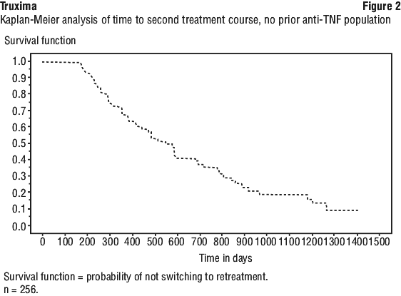
Depending on the circumstances of your disease or response to the drug, your doctor may decide to re-treat your RA with an additional course of Truxima®.

The dose of Truxima® for GPA and MPA patients is 375 mg per square metre of body surface area once a week for 4 weeks. For GPA or MPA Truxima® should be used together with glucocorticoids.

Overdose

As Truxima® is given to you under the supervision of your doctor, it is very unlikely that you will receive too much. However, if you experience any side effects after being given Truxima®, tell your doctor immediately.

While you are receiving Truxima®

Things you must do

If you are a woman of child bearing potential, you must use effective contraceptive methods to prevent pregnancy during treatment with Truxima® and for 12 months after completing treatment.

Tell your doctor if you become pregnant while receiving Truxima®.

Tell all doctors, dentists and pharmacists who are treating you that you are receiving Truxima®.

Tell your partner or caregiver you are receiving Truxima® and ask them to tell you if they notice any changes in your movement or behaviour. If they notice any changes you should tell your doctor about them immediately. Your doctor may need to perform some tests and alter your treatment.

Be sure to keep all your appointments with your doctor so that your progress can be checked. Your doctor will perform regular blood tests.

Things you must not do

Do not breast feed your infant during treatment with Truxima®. It is not known whether Truxima® crosses into human milk.

Do not take any other medicines whether they require a prescription or not without first telling your doctor or consulting with a pharmacist.

Things to be careful of

Be careful driving or operating machinery until you know how Truxima® affects you. Truxima® generally does not cause any problems with your ability to drive or operate machinery. However, as with many other medicines, Truxima® may cause dizziness in some people.

Side effects

Tell your doctor or pharmacist as soon as possible if you do not feel well while you are receiving Truxima®.

Do not be alarmed by this list of possible side effects. You may not experience any of them. Truxima® helps many people with RA, GPA or MPA but it may have unwanted side effects.

All medicines can have side effects. Sometimes they are serious, most of the time they are not. You may need medical treatment if you get some of the side effects.

Ask your doctor or pharmacist to answer any questions you may have.

The following is a list of the more common side effects.

During or after an infusion

Tell your doctor if you notice any of the following during or after receiving an infusion (particularly during the first 2 hours of receiving the first infusion) and they worry you:

  • fever, chills and severe shivering (most likely to occur)
  • swelling of the tongue, face, lips, mouth or throat which may cause difficulty breathing or swallowing
  • itchy rash and/or pinkish, itchy swellings on the skin
  • difficulty breathing and/or shortness of breath
  • wheezing or coughing
  • dizziness or lightheadedness, especially on standing up
  • high blood pressure
  • tremor
  • nausea (feeling sick) or vomiting
  • headache
  • fatigue (feeling tired) and/or feeling weak
  • rhinitis (a runny nose)
  • flushing
  • fast heart beat
  • chest pain
  • stomach pain or discomfort
  • throat irritation

These side effects are temporary and less likely to occur after the first infusion.

Your doctor may recommend that you take medication to prevent pain or allergy before you receive your Truxima® infusion.

The following is a list of other side effects which may occur during treatment with Truxima®. Tell your doctor if you notice any of them and they worry you:

  • aching muscles, muscle tenderness or weakness, muscle spasms
  • indigestion
  • painful, swollen joints
  • severe headache
  • high cholesterol
  • insomnia
  • diarrhoea
  • pins and needles, or decreased feeling in the skin
  • infections e.g. urinary tract infections, colds or chest infections including pneumonia

Tell your doctor immediately or go to Accident and Emergency at your nearest hospital if you notice any of the following:

  • infections with fever, severe chills, sore throat or mouth ulcers
  • severe skin rash, itching, hives
  • swelling of the face, lips, mouth or throat which may cause difficulty in swallowing or breathing, swelling of the hands, feet or ankles
  • one or a combination of the following: severe shortness of breath, severe difficulty breathing, severe wheezing, severe coughing
  • severe stomach pain, nausea or vomiting
  • vision loss associated with headaches, confusion and seizures
  • one or a combination of the following: confusion, disorientation or memory loss, changes in the way you move, walk or talk, decreased strength or progressive weakness in your body, blurred or loss of vision
  • yellowing of skin and eyes, light coloured bowel motions, dark coloured urine
  • fever, headache and stiff neck, incoordination (ataxia), personality change, hallucinations, altered consciousness, seizures or coma – these may be due to a serious brain infection (enteroviral meningoencephalitis), which can be fatal.

These are serious side effects. You may need urgent medical attention. Serious side effects are rare.

This is not a complete list of all possible side effects. Your doctor or pharmacist has a more complete list. Others may occur in some people and there may be some side effects not yet known.

Tell your doctor if you notice anything else that is making you feel unwell.

Ask your doctor or pharmacist if you don’t understand anything in this list.

Do not be alarmed by this list of possible side effects. You may not experience any of them.

After receiving Truxima®

Storage

Truxima® should be stored in the pharmacy or on the hospital ward.

The concentrated solution for infusion should be kept in a refrigerator at 2-8°C. It should not be frozen.

Truxima® should be stored away from light.

Product Description

Availability

Truxima® is available as 100 mg/10 mL and 500 mg/50 mL single dose vials.

Truxima® comes in packs of two vials for the 100mg/10 mL presentation and packs of one vial for the 500 mg/50 mL presentation.

What Truxima® looks like

Truxima® is available as a clear, colourless, concentrated solution for intravenous infusion. It is diluted before infusion into a vein.

Ingredients

Truxima® contains the active ingredient rituximab (rch). Truxima® comes in two strength, 100 mg and 500 mg. Each vial of Truxima® also contains the following inactive ingredients:

  • sodium citrate
  • polysorbate 80
  • sodium chloride
  • sodium hydroxide and/or hydrochloric acid

Sponsor

Celltrion Healthcare Australia Pty Ltd
Suite 13.03, 31 Market Street,
Sydney 2000, Australia
1800 325 228

Please check with your pharmacist for the latest Consumer Medicine Information.

Australian Registration Numbers:

  • 100 mg vial: AUST R 285816
  • 500 mg vial: AUST R 285815

This leaflet was prepared on October 2023

Published by MIMS February 2024

BRAND INFORMATION

Brand name

Truxima

Active ingredient

Rituximab

Schedule

S4


1 Name of Medicine

Truxima rituximab (rch) Concentrate for solution for intravenous (IV) infusion.

2 Qualitative and Quantitative Composition

Active ingredient.

Truxima contains the active ingredient rituximab (rch).
Truxima is supplied at a concentration of 10 mg/mL in either 100 mg (10 mL) or 500 mg (50 mL) single-use glass vials.

Excipients.

For the full list of excipients, see Section 6.1 List of Excipients.

3 Pharmaceutical Form

Truxima concentrate for solution for IV infusion is a sterile, clear, colourless, preservative-free, concentrated solution.

4 Clinical Particulars

4.1 Therapeutic Indications

Non-Hodgkin's lymphoma.

Truxima is indicated for treatment of patients with:
CD20 positive, previously untreated, Stage III/IV follicular, B-cell non-Hodgkin's lymphoma;
CD20 positive, relapsed or refractory low grade or follicular, B-cell non-Hodgkin's lymphoma;
CD20 positive, diffuse large B-cell non-Hodgkin's lymphoma, in combination with chemotherapy.

Chronic lymphocytic leukaemia.

Truxima is indicated for the treatment of patients with CD20 positive chronic lymphocytic leukaemia (CLL) in combination with chemotherapy.

Rheumatoid arthritis.

Truxima (rituximab) in combination with methotrexate is indicated for the treatment of adult patients with severe, active rheumatoid arthritis who have had an inadequate response or intolerance to at least one tumour necrosis factor (TNF) inhibitor therapy.
Truxima has been shown to reduce the rate of progression of joint damage as measured by x-ray when given in combination with methotrexate.

Granulomatosis with polyangiitis (Wegener's) (GPA) and microscopic polyangiitis (MPA).

Truxima in combination with glucocorticoids is indicated for the induction of remission in patients with severely active Granulomatosis with polyangiitis (GPA, also known as Wegener's granulomatosis) and Microscopic polyangiitis (MPA). The efficacy and safety of retreatment with rituximab have not been established.

4.2 Dose and Method of Administration

Truxima intravenous formulation is not intended for subcutaneous (SC) administration.
Truxima may be administered in an outpatient setting. Truxima should be administered as an intravenous infusion in an environment where full resuscitation facilities are immediately available, and under the close supervision of an experienced healthcare professional.
Substitution by any other biological medicinal product requires the consent of the prescribing physician.

Dosage. Non-Hodgkin's lymphoma.

Premedication, consisting of an analgesic/antipyretic (such as paracetamol) and an antihistamine should always be administered 30 to 60 minutes before each infusion of Truxima. Premedication with glucocorticoids should also be considered, particularly if Truxima is not given in combination with steroid-containing chemotherapy.

Relapsed or refractory low grade or follicular non-Hodgkin's lymphoma.

The recommended dosage of Truxima when used in monotherapy is 375 mg/m2 administered as an intravenous infusion once weekly for four weeks.
The recommended dosage of Truxima when used in combination with CHOP chemotherapy is 375 mg/m2 administered on day 1 of each chemotherapy cycle (6 cycles).

Previously untreated stage III/IV follicular non-Hodgkin's lymphoma.

The recommended dosage of Truxima in combination with chemotherapy is 375 mg/m2 administered on day 1 of each chemotherapy cycle for up to 8 cycles as induction therapy.
Truxima should be administered prior to the administration of chemotherapy. Any infusion related reactions should have settled before chemotherapy is instituted.

Maintenance treatment in follicular lymphoma.

Previously untreated patients who have responded to induction treatment may receive maintenance therapy with Truxima given at 375 mg/m2 body surface area once every 2 months until disease progression or for a maximum period of two years.
Relapsed/refractory patients who have responded to induction treatment may receive maintenance therapy with Truxima given at 375 mg/m2 body surface area once every 3 months until disease progression or for a maximum period of two years.

Diffuse large B-cell non-Hodgkin's lymphoma.

The recommended dosage for Truxima in combination with CHOP chemotherapy is 375 mg/m2, administered as an intravenous infusion on day 1 of each chemotherapy cycle, for up to 8 cycles.

Chronic lymphocytic leukaemia.

Premedication, consisting of an analgesic/antipyretic such as paracetamol and an antihistamine should always be administered 30 to 60 minutes before each infusion of Truxima. Premedication with glucocorticoids should also be considered, particularly if Truxima is not given in combination with steroid-containing chemotherapy.
The recommended dosage of rituximab in combination with chemotherapy is 375 mg/m2 administered on day 1 of the first treatment cycle followed by 500 mg/m2 administered on day 1 of each subsequent cycle, for a total of 6 cycles (see Section 5 Pharmacological Properties, Clinical trials). The chemotherapy should be given after the infusion of Truxima.
Prophylaxis with adequate hydration and administration of uricostatics starting 48 hours prior to the start of therapy is recommended for CLL patients to reduce the risk of tumour lysis syndrome. For CLL patients whose lymphocyte counts are > 25 x 109/L it is recommended to administer methylprednisolone 100 mg IV shortly before infusion with Truxima to decrease the rate and severity of acute infusion reactions and/or cytokine release syndrome.
Dosage adjustments during treatment. No dose reductions of Truxima are recommended. When Truxima is given in combination with chemotherapy, standard dose reductions for the chemotherapeutic drugs should be applied.

First infusion.

The recommended initial rate of infusion is 50 mg/h. If hypersensitivity or infusion-related events do not occur, escalate the infusion rate in 50 mg/h increments every 30 minutes, to a maximum of 400 mg/h. If hypersensitivity or an infusion-related event develops, the infusion should be temporarily slowed or interrupted (see Section 4.4 Special Warnings and Precautions for Use). The infusion can continue at one-half the previous rate upon improvement of patient symptoms.

Subsequent infusions.

Subsequent Truxima infusions can be administered at an initial rate of 100 mg/h and increased by 100 mg/h increments at 30-minute intervals, to a maximum of 400 mg/h.

Rheumatoid arthritis.

Premedication consisting of an analgesic/antipyretic such as paracetamol and an antihistamine should always be administered 30 to 60 minutes before each infusion of Truxima.
Premedication with glucocorticoids should also be administered in order to reduce the frequency and severity of IRRs. Patients should receive 100 mg IV methylprednisolone to be completed 30 minutes prior to each Truxima infusion (see Section 4.4 Special Warnings and Precautions for Use, Rheumatoid arthritis (RA), granulomatosis with polyangiitis (Wegener's) (GPA) and microscopic polyangiitis (MPA)).
A course of Truxima consists of two 1000 mg IV infusions. The recommended dosage of Truxima is 1000 mg by IV infusion followed by a second 1000 mg IV infusion two weeks later. The course of Truxima is given concomitantly with the dose of MTX tolerated by the patient. The minimal effective dose is not yet known.
Background therapy with glucocorticoids, salicylates, nonsteroidal anti-inflammatory drugs, or analgesics can be continued during treatment with Truxima.
Disease activity should be regularly monitored. Patients may receive further courses of treatment, based on signs and symptoms of disease. In clinical studies, no patient received a second course of rituximab treatment within 16 weeks of the first infusion of the first course. The time interval between courses was variable, with the majority of patients who received additional courses doing so 6 -12 months after the previous course. Some patients required even less frequent retreatment. The efficacy and safety of further courses is comparable to the first course.
Human anti chimeric antibodies (HACA) develop in some patients after the first course of rituximab. The presence of HACA may be associated with the worsening of infusion or allergic reactions after the second infusion of subsequent course. Furthermore, in one case with HACA, failure to deplete B-cells after receipt of further treatment courses has been observed. Thus, the benefit/risk balance of therapy with Truxima should be carefully considered before administering subsequent courses of Truxima. If a repeat course of treatment is considered, it should not be given at an interval less than 16 weeks.
Dosage adjustments during treatment.

First infusion of each course.

The recommended initial rate for infusion is 50 mg/h; after the first 30 minutes, it can be escalated in 50 mg/h increments every 30 minutes, to a maximum of 400 mg/h.

Second infusion of each course.

Subsequent doses of Truxima can be infused at an initial rate of 100 mg/h, and increased by 100 mg/h increments at 30 minutes intervals, to a maximum of 400 mg/h.
Rheumatoid arthritis only; alternative subsequent, faster, infusion schedule. In RA, with a dose of 1000 mg Truxima, if there are no infusion related reactions or other reasons to slow or cease the infusion, the standard infusion schedules shown above result in an estimated duration of infusion of 4 h 15 minutes for the first infusion and 3 h 15 minutes for the second infusion in each course.
If patients do not experience a serious infusion related reaction with their first or subsequent infusions administered over the standard infusion schedule, a more rapid infusion can be administered for second and subsequent infusions using a concentration of 4 mg/mL in a 250 mL volume. Initiate at a rate of 250 mg/h for the first 30 minutes and then 600 mg/h for the next 90 minutes. With this infusion schedule, the 1000 mg/250 mL infusion will generally be completed in 2 h.
Patients who have clinically significant cardiovascular disease, including arrhythmias, or previous serious infusion reactions to any prior biologic therapy or to rituximab, should not be administered the more rapid, 2 h, infusion.

Granulomatosis with polyangiitis (Wegener's) (GPA) and microscopic polyangiitis (MPA).

Premedication consisting of an analgesic/antipyretic such as paracetamol and an antihistamine should always be administered 30 to 60 minutes before each infusion of Truxima.
The recommended dosage of Truxima for treatment of GPA and MPA is 375 mg/m2 body surface area, administered as an IV infusion once weekly for 4 weeks.
Methylprednisolone 1000 mg IV per day for 1 to 3 days is recommended in combination with Truxima to treat severe vasculitis symptoms, followed by oral prednisone 1 mg/kg/day (not to exceed 80 mg/day, and tapered as rapidly as possible per clinical need) during and after Truxima treatment.

First infusion.

The recommended initial infusion rate for Truxima is 50 mg/h; subsequently the rate can be escalated in 50 mg/h increments every 30 minutes to a maximum of 400 mg/h.

Subsequent infusions.

Subsequent infusions of Truxima can be administered at a rate of 100 mg/h and increased by 100 mg/h increments every 30 minutes to a maximum of 400 mg/h.
Pneumocystis jiroveci pneumonia (PJP) prophylaxis is recommended for patients with GPA and MPA during and following Truxima treatment, as appropriate.

Special populations.

Elderly.

No dose adjustment is required in elderly patients (aged > 65 years).

Preparation.

Truxima vials do not contain an antimicrobial agent or preservative; therefore, care must be taken to ensure the sterility of the vials and prepared solution. Product is for single use in one patient only. Discard any residue.
Aseptically withdraw the necessary amount of Truxima and dilute to a calculated concentration between 1 mg/mL to 4 mg/mL of rituximab into an infusion bag containing either 0.9% sodium chloride or 5% dextrose in water.To mix the solution, gently invert the bag to avoid foaming. Parenteral drug products should be inspected visually for particulate matter and discolouration prior to administration.
To reduce microbiological hazard, prepared infusion solutions of Truxima should be used as soon as practicable after dilution. If necessary, the prepared solutions may be stored in the refrigerator (2℃ to 8℃) for up to 24 hours. This timeframe allows for the temporary interruption of the infusion and subsequent recommencement if the patient has an infusion reaction (see Method of administration).
No incompatibilities between Truxima and polyvinyl chloride or polyethylene bags have been observed.

Method of administration.

The Truxima solution for infusion should be administered intravenously through a dedicated line.
As with all parenteral products, appropriate aseptic technique should be used during the administration of Truxima. Do not administer as an intravenous push or bolus. Hypersensitivity reactions may occur whenever protein solutions such as Truxima are administered (see Section 4.4 Special Warnings and Precautions for Use).

4.3 Contraindications

Truxima is contraindicated in patients with known hypersensitivity to rituximab, to any of its excipients or to murine proteins.

4.4 Special Warnings and Precautions for Use

Progressive multifocal leukoencephalopathy (PML).

Use of Truxima may be associated with an increased risk of progressive multifocal leukoencephalopathy (PML). Patients must be monitored for any new or worsening neurological symptoms or signs suggestive of PML. Physicians treating patients should consider PML in the differential diagnosis of patients reporting neurological symptoms and consultation with a neurologist should be considered as clinically indicated.
Physicians should be particularly alert to symptoms suggestive of PML that the patient may not notice (e.g. cognitive, neurological or psychiatric symptoms). If such symptoms occur, further administration of Truxima should be immediately suspended until a diagnosis of PML has been excluded. To establish or exclude a diagnosis of PML evaluation including MRI scan, CSF testing for JC viral DNA and repeat neurological assessments, should be considered. Once PML has been excluded, the administration of Truxima may resume.
If a diagnosis of PML is confirmed Truxima must be permanently discontinued. Patients should also be advised to inform their partner or caregivers about their treatment, since they may notice symptoms that the patient is not aware of.

Non-Hodgkin's lymphoma and chronic lymphocytic leukaemia.

Infusion-related reactions.

Rituximab is associated with infusion-related reactions, which may be related to release of cytokines and/or other chemical mediators. Cytokine release syndrome may be indistinguishable from acute hypersensitivity reactions.

Infusion-related reactions for rituximab.

Severe infusion-related reactions with fatal outcome have been reported during post-marketing use. Severe reactions usually manifested within 30 minutes to 2 hours after starting the first rituximab infusion, were characterised by pulmonary events and included, in some cases, rapid tumour lysis and features of tumour lysis syndrome in addition to fever, chills, rigors, hypotension, urticaria, angio-oedema and other symptoms. Patients with a high tumour burden or with a high number (> 25 x 109/L) of circulating malignant cells such as patients with chronic lymphocytic leukaemia (CLL) and mantle cell lymphoma may be at higher risk of developing severe infusion-related reactions. Infusion reaction symptoms are usually reversible with interruption of the infusion. Treatment of infusion-related symptoms with an antihistamine and an analgesic/antipyretic (such as paracetamol) is recommended. Additional treatment with bronchodilators or IV saline may be indicated. In most cases, the infusion can be resumed at a 50% reduction in rate (e.g. from 100 mg/h to 50 mg/h) when symptoms have completely resolved. Most patients who have experienced non-life threatening infusion-related reactions have been able to complete the full course of rituximab therapy. Further treatment of patients after complete resolution of signs and symptoms has rarely resulted in repeated severe infusion-related reactions.
Patients with a high number (> 25 x 109/L) of circulating malignant cells or high tumour burden such as patients with CLL and mantle cell lymphoma, who may be at higher risk of especially severe infusion-related reactions, should only be treated with extreme caution and when other therapeutic alternatives have been exhausted. These patients should be very closely monitored throughout the first infusion. Consideration should be given to the use of a reduced infusion rate for the first infusion in these patients, or a split dosing over two days during the first cycle and any subsequent cycles if the lymphocyte count is still > 25 x 109/L.

Hypersensitivity reactions/anaphylaxis.

Anaphylactic and other hypersensitivity reactions have been reported following the intravenous administration of proteins to patients. Adrenaline, antihistamines and corticosteroids should be available for immediate use in the event of a hypersensitivity reaction to Truxima.

Pulmonary events.

Pulmonary events have included hypoxia, lung infiltration, and acute respiratory failure. Some of these events have been preceded by severe bronchospasm and dyspnoea. In some cases, symptoms worsened over time, while in others initial improvement was followed by clinical deterioration. Therefore, patients experiencing pulmonary events or other severe infusion-related symptoms should be closely monitored until complete resolution of their symptoms occurs. Patients with a history of pulmonary insufficiency or those with pulmonary tumour infiltration may be at greater risk of poor outcome and should be treated with increased caution. Acute respiratory failure may be accompanied by events such as pulmonary interstitial infiltration or oedema, visible on a chest x-ray. The syndrome usually manifests itself within one or two hours of initiating the first IV infusion. Patients who experience severe pulmonary events should have their infusion interrupted immediately and should receive aggressive symptomatic treatment. Since initial improvement of clinical symptoms may be followed by deterioration, these patients should be closely monitored until the pulmonary event has resolved.

Rapid tumour lysis.

Truxima mediates the rapid lysis of benign and malignant CD20-positive cells. Signs and symptoms (e.g. hyperuricaemia, hyperkalaemia, hypocalcaemia, acute renal failure, elevated LDH) consistent with tumour lysis syndrome (TLS) have been reported to occur after the first rituximab infusion in patients with high numbers of circulating malignant lymphocytes. Prophylaxis for TLS should be considered for patients at risk of developing rapid tumour lysis (e.g. patients with a high tumour burden or with a high number (> 25 x 109/L) of circulating malignant cells such as patients with CLL and mantle cell lymphoma). These patients should be followed closely and appropriate laboratory monitoring performed. Appropriate medical therapy should be provided for patients who develop signs and symptoms consistent with rapid tumour lysis. Following treatment for and complete resolution of signs and symptoms, subsequent rituximab therapy has been administered in conjunction with prophylactic therapy for TLS in a limited number of cases.

Cardiovascular.

Since hypotension may occur during rituximab infusion, consideration should be given to withholding antihypertensive medications 12 hours prior to and throughout rituximab infusion. Angina pectoris or cardiac arrhythmia, such as atrial flutter and fibrillation, heart failure or myocardial infarction have occurred in patients treated with rituximab. Therefore, patients with a history of cardiac disease should be monitored closely. Infusions should be discontinued in the event of serious or life-threatening cardiac arrhythmias.

Monitoring of blood counts.

Although rituximab is not myelosuppressive in monotherapy, caution should be exercised when considering treatment of patients with neutrophil counts of < 1.5 x 10⁹/L and/or platelet counts of < 75 x 109/L, as clinical experience with such patients is limited. Rituximab has been used in patients who underwent autologous bone marrow transplantation and in other risk groups with a presumable reduced bone marrow function without inducing myelotoxicity.
Consideration should be given to the need for regular full blood counts, including platelet counts, during monotherapy with rituximab. When rituximab is given in combination with CHOP or CVP chemotherapy, regular full blood counts should be performed according to usual medical practice.

Infections.

Truxima treatment should not be initiated in patients with severe active infections.
Cases of Hepatitis B virus (HBV) reactivation, occasionally with fulminant hepatitis, hepatic failure, and death have been reported in some patients with haematologic malignancies treated with rituximab. The majority of patients received rituximab in combination with chemotherapy. Isolated cases have been reported in patients who either had evidence of antibodies against Hepatitis B surface antigen before treatment or did not have any such antibodies. The median time to diagnosis of hepatitis was approximately 4 months after the initiation of rituximab and approximately one month after the last dose. Analysis of events revealed rituximab use has been associated with hepatitis B (HB) reactivation in patients with positive HB surface antigen (HBsAg +ve) as well as negative HB surface antigen and positive anti-HB core antibody (HBsAg -ve/HBcAb +ve), particularly when administered in combination with steroids or chemotherapy.
HBV screening should be performed in all patients before initiation of treatment with Truxima. At a minimum this should include HBsAg-status and HBcAb-status. These can be complemented with other appropriate markers as per local guidelines. Patients with active HB disease should not be treated with Truxima. Reactivation of HBV infection is a well-known complication in patients with chronic hepatitis B, especially in those receiving cytotoxic or immunosuppressive therapy. In addition, non-Hodgkin's lymphoma of itself may be an independent risk factor for HBV reactivation. Patients with positive HB serology should consult a liver disease specialist before the start of treatment and should be monitored and managed according to guidelines to prevent HB reactivation.
In patients who develop reactivation of viral hepatitis B, Truxima and any concomitant chemotherapy should be discontinued and appropriate treatment including antiviral therapy initiated. There are insufficient data regarding the safety of resuming therapy with rituximab in patients who develop hepatitis subsequent to HBV reactivation.
The following additional serious viral infections, either new, reactivated or exacerbated, have been identified in clinical studies or post-marketing reports. The majority of patients were profoundly immune-suppressed. These viral infections included JC virus [progressive multifocal leukoencephalopathy (PML)], cytomegalovirus, herpes simplex virus, parvovirus B19, varicella zoster virus, West Nile virus and hepatitis C. In some cases, the viral infections occurred up to one year following discontinuation of rituximab and have resulted in death.

Enteroviral meningoencephalitis.

Cases of enteroviral meningoencephalitis, including fatal cases, have been reported following use of rituximab. Physicians should consider enteroviral meningoencephalitis in the differential diagnosis of patients reporting symptoms such as fever, headache, stiff neck, ataxia, psychiatric symptoms, seizures, altered consciousness or coma. A high level of suspicion is required to facilitate early diagnosis and appropriate treatment and to minimise the risk of severe outcomes.

Skin reactions.

Severe skin reactions such as toxic epidermal necrolysis and Stevens-Johnson syndrome, some with fatal outcome, have been reported (see Section 4.8 Adverse Effects (Undesirable Effects), Post-marketing experience, Non-Hodgkin's lymphoma and chronic lymphocytic leukaemia). In case of such an event, with a suspected relationship to rituximab, treatment should be permanently discontinued.

Immunisation.

The safety of immunisation with live viral vaccines, following rituximab therapy has not been studied and vaccination with live virus vaccines is not recommended.
Patients treated with Truxima may receive non-live vaccinations. However, with non-live vaccines response rates may be reduced. In a non-randomised study, patients with relapsed low-grade NHL who received rituximab monotherapy when compared to healthy untreated controls had a lower rate of response to vaccination with tetanus recall antigen (16% vs. 81%) and Keyhole Limpet Haemocyanin (KLH) neoantigen (4% vs. 76% when assessed for > 2-fold increase in antibody titer).
Mean pre-therapeutic antibody titers against a panel of antigens (Streptococcus pneumoniae, influenza A, mumps, rubella, varicella) were maintained for at least 6 months after treatment with rituximab.

Progressive multifocal leukoencephalopathy (PML).

Cases of progressive multifocal leukoencephalopathy (PML) have been reported during use of rituximab in NHL and CLL. The majority of patients had received rituximab in combination with chemotherapy or as part of a haematopoietic stem cell transplant. (See Boxed Warnings; Section 4.8 Adverse Effects (Undesirable Effects), Post-marketing experience).

Rheumatoid arthritis (RA), granulomatosis with polyangiitis (Wegener's) (GPA) and microscopic polyangiitis (MPA).

The efficacy and safety of rituximab for the treatment of autoimmune diseases other than RA, GPA and MPA have not been established.
Truxima in combination with glucocorticoids is indicated for the induction of remission in patients with severely active GPA and MPA. Limited data are available on the efficacy and safety of subsequent courses of rituximab in patients with GPA and MPA, hence, the efficacy and safety of retreatment with rituximab for these diseases have not been established.
Limited data from retrospective experience may suggest that vasculitis versus granulomatous disease manifestations of GPA have a differential treatment response to rituximab. These reports lack confirmatory data. Most available reports, including controlled studies, clearly show that both vasculitic and granulomatous disease manifestations of GPA respond well to rituximab.

Infusion-related reactions.

Rituximab is associated with infusion-related reactions (IRRs), which may be related to release of cytokines and/or other chemical mediators. Premedication consisting of an analgesic/antipyretic drug and an antihistamine drug should always be administered before each infusion of Truxima. For RA patients, premedication with glucocorticoids should also be administered before each infusion of Truxima in order to reduce the frequency and severity of IRRs (see Section 4.8 Adverse Effects (Undesirable Effects), Experience from clinical trials in rheumatoid arthritis; Section 4.2 Dose and Method of Administration, Rheumatoid arthritis).
For RA patients, most IRRs reported in clinical trials were mild to moderate in severity.
Severe IRRs with fatal outcome have been reported in the post-marketing setting (see Section 4.8 Adverse Effects (Undesirable Effects), Post-marketing experience, Rheumatoid arthritis, granulomatosis with polyangiitis (Wegener's) (GPA) and microscopic polyangiitis (MPA) patients). Closely monitor patients with pre-existing cardiac conditions and those who experienced prior cardiopulmonary adverse reactions. The most common symptoms were headache, pruritus, throat irritation, flushing, rash, urticaria, hypertension, and pyrexia. In general, the proportion of patients experiencing any infusion reaction was higher following the first infusion of any treatment course than following the second infusion. Subsequent rituximab infusions were better tolerated by patients than the initial infusion. Fewer than 1% of patients experienced serious IRRs, with most of these reported during the first infusion of the first course (see Section 4.8 Adverse Effects (Undesirable Effects), Experience from clinical trials in rheumatoid arthritis). The reactions reported were usually reversible with a reduction in rate, or interruption, of rituximab infusion and administration of an anti-pyretic, an antihistamine, and, occasionally, oxygen, IV saline or bronchodilators, and glucocorticoids if required. Depending on the severity of the IRR and the required interventions, temporarily or permanently discontinue Truxima. In most cases, the infusion can be resumed at a 50% reduction in rate (e.g. from 100 mg/h to 50 mg/h) when symptoms have completely resolved.
For GPA and MPA patients, IRRs were similar to those seen for RA patients in clinical trials (see Section 4.8 Adverse Effects (Undesirable Effects), Clinical trial experience in granulomatosis with polyangiitis (Wegener's) (GPA) and microscopic polyangiitis (MPA)). For GPA and MPA patients, rituximab was given in combination with high doses of glucocorticoids (see Section 4.2 Dose and Method of Administration), which may reduce the incidence and severity of these events (see information for RA indication above).

Hypersensitivity reactions/anaphylaxis.

Anaphylactic and other hypersensitivity reactions have been reported following the intravenous administration of proteins to patients. Medicinal products for the treatment of hypersensitivity reactions, e.g. adrenaline, antihistamines and glucocorticoids, should be available for immediate use in the event of an allergic reaction during administration of Truxima. The presence of HACA may be associated with worsening infusion or allergic reactions after the second infusion of subsequent courses.

Infections.

Serious infections, including fatalities, can occur during therapy with rituximab. Based on the mechanism of action of rituximab and the knowledge that B cells play an important role in maintaining normal immune response, patients may have an increased risk of infection following rituximab therapy (see Section 5.1 Pharmacodynamic Properties). A small proportion of patients (approximately 8%) had prolonged peripheral B-cell depletion (< 80 cells/microL) lasting 2 years or more after their last dose of rituximab. Approximately a third of these patients had low B-cell counts (< 80 cells/microL) prior to starting rituximab treatment.
Truxima should not be administered to patients with an active infection or severely immunocompromised patients (e.g. where levels of CD4 or CD8 are very low). Physicians should exercise caution when considering the use of rituximab in patients with a history of recurring or chronic infections or with underlying conditions which may further predispose patients to serious infection. Patients who develop infection following rituximab therapy should be promptly evaluated and treated appropriately.
Cases of reactivation of hepatitis B infection, including those with a fatal outcome, have been reported in RA, GPA and MPA patients receiving rituximab. Analysis of events revealed rituximab use has been associated with hepatitis B (HB) reactivation in patients with positive HB surface antigen (HBsAg +ve) as well as negative HB surface antigen and positive anti-HB core antibody (HBsAg -ve/HBcAb +ve), particularly when administered in combination with steroids.
Hepatitis B virus (HBV) screening should be performed in all patients before initiation of treatment with rituximab. At a minimum this should include HBsAg-status and HBcAb-status. These can be complemented with other appropriate markers as per local guidelines. Patients with active HB disease should not be treated with rituximab. Patients with positive HB serology should consult a liver disease specialist before the start of treatment and should be monitored and managed according to guidelines to prevent HB reactivation.

Enteroviral meningoencephalitis.

Cases of enteroviral meningoencephalitis, including fatal cases, have been reported following use of rituximab. Physicians should consider enteroviral meningoencephalitis in the differential diagnosis of patients reporting symptoms such as fever, headache, stiff neck, ataxia, psychiatric symptoms, seizures, altered consciousness or coma. A high level of suspicion is required to facilitate early diagnosis and appropriate treatment and to minimise the risk of severe outcomes.

Skin reactions.

Severe skin reactions such as toxic epidermal necrolysis and Stevens-Johnson syndrome, some with fatal outcome, have been reported (see Section 4.8 Adverse Effects (Undesirable Effects), Post-marketing experience, Rheumatoid arthritis, granulomatosis with polyangiitis (Wegener's) (GPA) and microscopic polyangiitis (MPA) patients). In case of such an event, with a suspected relationship to rituximab, treatment should be permanently discontinued. Re-administration must be carefully assessed based on the individual patient's benefit-risk profile.

Progressive multifocal leukoencephalopathy (PML).

Cases of progressive multifocal leukoencephalopathy (PML) have been reported following use of rituximab for the treatment of autoimmune diseases including RA and GPA. Several but not all of the reported cases involved patients with recognised risk factors for PML, including the underlying disease and long term immunosuppressive therapy or chemotherapy. (See Boxed Warnings; Section 4.4 Special Warnings and Precautions for Use). The efficacy and safety of rituximab for the treatment of autoimmune diseases other than RA, GPA and MPA has not been established.

Immunisation.

For RA patients, physicians should review the patient's vaccination status and follow current immunisation guidelines prior to treatment with rituximab. Vaccination should be completed at least 4 weeks prior to first administration of rituximab.
The safety of immunisation with live viral vaccines following rituximab therapy has not been studied. Therefore, vaccination with live virus vaccines is not recommended whilst on rituximab or whilst peripherally B cell depleted.
Patients treated with rituximab may receive non-live vaccinations. However, response rates to non-live vaccines may be reduced. In a randomised study, patients with RA treated with rituximab and MTX had comparable response rates to tetanus recall antigen (39% vs. 42%), reduced rates to pneumococcal polysaccharide vaccine (43% vs. 82% to at least 2 pneumococcal antibody serotypes), and KLH neoantigen (34% vs. 80%), when given at least 6 months after rituximab as compared to patients only receiving MTX. Should non-live vaccinations be required whilst receiving rituximab therapy, these should be completed at least 4 weeks prior to commencing the next course of rituximab.
In the overall experience of rituximab repeat treatment in RA patients over one year, the proportions of patients with positive antibody titers against S. pneumoniae, influenza, mumps, rubella, varicella and tetanus toxoid were generally similar to the proportions at baseline.

Cardiovascular events.

Patients with a history of cardiac disease should be monitored closely during infusions. Infusions should be discontinued in the event of serious or life-threatening cardiac arrhythmias (see Section 4.4 Special Warnings and Precautions for Use, Non-Hodgkin's lymphoma and chronic lymphocytic leukaemia, Cardiovascular). There are no data on the safety of rituximab in patients with moderate or severe heart failure (NYHA class III or IV) or severe, uncontrolled cardiovascular disease. In patients treated with rituximab, the occurrence of pre-existing ischaemic cardiac conditions becoming symptomatic, such as angina pectoris, has been observed, as well as cardiac arrhythmias such as atrial fibrillation and flutter. Therefore, in patients with a known cardiac history, the risk of cardiovascular complications resulting from infusion reactions should be considered before treatment with rituximab and patients monitored closely (see Section 4.4 Special Warnings and Precautions for Use, Infusion-related reactions). Since hypotension may occur during rituximab infusion, consideration should be given to withholding anti-hypertensive medications 12 hours prior to the rituximab infusion.

Methotrexate (MTX) naïve populations with RA.

The use of rituximab is not recommended in MTX-naïve patients since a favourable benefit-risk relationship has not been established.

Use in GPA or MPA patients aged ≥ 65 years.

In the RAVE study, GPA or MPA patients aged ≥ 65 years in both treatment groups demonstrated an increased risk of SAEs, ≥ Grade 3 AEs, death or hospitalisation. It is recommended that patients ≥ 65 years receiving treatment for GPA or MPA are monitored closely (see Section 4.8 Adverse Effects (Undesirable Effects)).

Concomitant/sequential use of other DMARDs in RA.

The concomitant use of rituximab and antirheumatic therapies other than those specified under the RA indication and dosing is not recommended.
Limited data are available on the safety of the use of biologic agents or DMARDs other than MTX in patients exhibiting peripheral B cell depletion following treatment with rituximab. If biologic agents and/or DMARDs are used following rituximab therapy, patients should be observed for signs of infection.

Malignancy.

Immunomodulatory drugs may increase the risk of malignancy. On the basis of limited experience with rituximab in RA patients (see Section 4.8 Adverse Effects (Undesirable Effects), Experience from clinical trials in rheumatoid arthritis) a possible risk for the development of solid tumours cannot be excluded at this time, although present data do not seem to suggest any increased risk.

Use in renal or hepatic impairment.

The safety and effectiveness of rituximab in patients with renal or hepatic impairment has not been established. MTX is contraindicated in such patients and since rituximab is given in combination with MTX, these patients were not included in the clinical studies for RA.
In the RAVE study, significant renal impairment was associated with an increased risk of experiencing any adverse event including SAEs, infections, hospitalisation or death. This increased risk was regardless of treatment strategy (rituximab or CYC). It is recommended that patients with significant renal impairment receiving treatment for GPA or MPA are monitored closely (see Section 4.8 Adverse Effects (Undesirable Effects)).

General precautions.

Traceability.

In order to improve the traceability of biological medicines, the trade name and the batch number of the administered product should be clearly recorded in the patient's medical record and/or dispensing record.

Paediatric use.

The safety and effectiveness of rituximab in paediatric patients have not been established. Hypogammaglobulinaemia has been observed in paediatric patients treated with rituximab, in some cases severe and requiring long-term immunoglobulin substitution therapy. The consequences of long term B cell depletion in paediatric patients are unknown.

Use in the elderly.

See Section 4.2 Dose and Method of Administration, Special populations; Section 4.8 Adverse Effects (Undesirable Effects), Subpopulations.

Effects on laboratory tests.

See Section 4.8 Adverse Effects (Undesirable Effects), Laboratory abnormalities.

4.5 Interactions with Other Medicines and Other Forms of Interactions

Currently, there are limited data on possible drug interactions with rituximab.
In CLL patients, co-administration with intravenous rituximab did not appear to have an effect on the pharmacokinetics of fludarabine or cyclophosphamide. In addition, there was no apparent effect of fludarabine and cyclophosphamide on the pharmacokinetics of rituximab.
Co-administration with MTX had no effect on the pharmacokinetics of intravenous rituximab in RA patients.
Patients with human anti-mouse antibody or human anti-chimeric antibody (HAMA/HACA) titres may have allergic or hypersensitivity reactions when treated with other diagnostic or therapeutic monoclonal antibodies.
The tolerability of simultaneously or sequential combination of rituximab with chemotherapy other than CHOP or CVP, or agents which are liable to cause depletion of normal B cells is not well defined.
In a small cohort of patients with RA, 110 patients received subsequent therapy with other DMARDs (including biologicals). Patients received subsequent DMARDs 4-6 months following therapy with rituximab and generally while peripherally B cell depleted. The rate of clinically relevant infections was 7.8 per 100 patient years.

4.6 Fertility, Pregnancy and Lactation

Effects on fertility.

No animal studies have been performed to determine the effects of rituximab on fertility in males or females.
(Category C)
It is not known whether rituximab can cause foetal harm when administered to a pregnant woman. There are no adequate and well-controlled data from studies in pregnant women, however transient B-cell depletion and lymphocytopenia have been reported in some infants born to mothers exposed to rituximab. In clinical studies in patients with RA, three pregnancies occurred following exposure to rituximab + MTX with two resulting in spontaneous abortions and the third ongoing at the time. Rituximab has been shown to cause B-cell depletion in the monkey foetus. Rituximab should not be given to a pregnant woman, unless the potential benefit outweighs the potential risk.
Women of child-bearing potential must use effective contraceptive methods during treatment and for 12 months following rituximab therapy.
Developmental toxicity studies performed in cynomolgus monkeys revealed no evidence of embryotoxicity in utero at relative exposure levels (AUC) similar to that anticipated clinically. New born offspring of maternal animals exposed to rituximab during lactation and/or gestation showed no untoward toxicity except for depleted B cell populations during the post-natal phase at the same relative exposure. B cell levels in human neonates following maternal exposure to rituximab have not been studied.
It is not known whether rituximab is excreted in human milk. In monkey studies, rituximab was excreted in the milk and was detected in the serum of breast-fed infants. Reversible B-cell depletion was observed in all monkey infants exposed to rituximab via maternal transfer during lactation and/or gestation. It is recommended that a nursing woman discontinue breastfeeding whilst undergoing treatment with rituximab.

4.7 Effects on Ability to Drive and Use Machines

It is not known whether rituximab has an effect on the ability to drive and operate machines, though the pharmacologic activity and adverse events reported to date do not indicate that such an effect is to be expected.

4.8 Adverse Effects (Undesirable Effects)

Experience from clinical trials in haemato-oncology.

The most common adverse reactions of rituximab (incidence ≥ 25%) observed in patients with NHL are infusion-related reactions, fever, chills, infection, asthenia and lymphopenia. The most important serious adverse reactions of rituximab are infusion-related reactions, tumour lysis syndrome, mucocutaneous toxicities, hepatitis B reactivation with fulminant hepatitis, PML, other viral infections, cardiac arrhythmias, renal toxicity, and bowel obstruction and perforation.
The frequencies of adverse drug reactions (ADRs) reported with rituximab alone or in combination with chemotherapy are summarised in Tables 1 and 2 and are based on data from clinical trials. These ADRs had either occurred in single arm studies or had occurred with at least a 2% difference compared to the control arm in at least one of the major randomised clinical trials. ADRs are added to the appropriate category in Tables 1 and 2 according to the highest incidence seen in any of the major clinical trials. Within each frequency grouping ADRs are listed in descending order of severity. Frequencies are defined as very common ≥ 1/10 (≥ 10%), common ≥ 1/100 to < 1/10 (≥ 1% to < 10%) and uncommon ≥ 1/1,000 to < 1/100 (≥ 0.1% to < 1%).
Rituximab monotherapy/maintenance therapy. The ADRs in Table 1 are based on data from single-arm studies including 356 patients with low-grade or follicular lymphoma, treated with rituximab weekly as a single agent for the treatment or re-treatment of non-Hodgkin's lymphoma (see Section 5 Pharmacological Properties, Clinical trials). Table 1 also contains ADRs based on data from 671 patients with follicular lymphoma who received rituximab as maintenance therapy for up to 2 years following response to initial induction with CHOP, R-CHOP, R-CVP or R-FCM (see Section 5 Pharmacological Properties, Clinical trials). The ADRs were reported up to 12 months after treatment with monotherapy and up to 1 month after treatment with rituximab maintenance.
Rituximab in combination with chemotherapy in NHL and CLL. The ADRs listed in Table 2 are based on rituximab-arm data from controlled clinical trials that occurred in addition to those seen with monotherapy/maintenance therapy and/or at a higher frequency grouping: 202 patients with diffuse large B-cell lymphoma (DLBCL) treated with R-CHOP, from 234 and 162 patients with follicular lymphoma treated with R-CHOP or R-CVP, respectively, and from 397 previously untreated CLL patients and 274 relapsed/refractory CLL patients treated with rituximab in combination with fludarabine and cyclophosphamide (R-FC) (see Section 5 Pharmacological Properties, Clinical trials).
The safety information of rituximab in combination with certain chemotherapy regimens is limited. When rituximab is used with other chemotherapy medicines, prescribers are advised to consider the adverse reaction profile of the component medicine(s).
The following terms have been reported as adverse events, however, were reported at a similar (< 2% difference between the groups) or lower incidence in the rituximab-arms compared to control arms: haematotoxicity, neutropenic infection, urinary tract infection, septic shock, superinfection lung, implant infection, septicaemia staphylococcal, lung infection, rhinorrhoea, pulmonary oedema, cardiac failure, sensory disturbance, venous thrombosis, mucosal inflammation nos, influenza-like illness, oedema lower limb, abnormal ejection fraction, pyrexia, general physical health deterioration, fall, multi-organ failure, venous thrombosis deep limb, positive blood culture, diabetes mellitus inadequate control.

Further information on selected, serious adverse drug reactions.

Infusion-related reactions.

Monotherapy - 4 weeks treatment.

Signs and symptoms suggestive of an IRR were reported in more than 50% of patients in clinical trials, and were predominantly seen during the first infusion. Hypotension, fever, chills, rigors, urticaria, bronchospasm, sensation of tongue or throat swelling (angioedema), nausea, fatigue, headache, pruritus, dyspnea, rhinitis, vomiting, flushing, and pain at disease sites have occurred in association with rituximab infusion as part of an infusion-related symptom complex. Some features of TLS have also been observed.

Maintenance treatment (NHL) up to 2 years.

Non-serious signs and symptoms suggestive of an infusion-related reaction were reported in 41% of patients for general disorders (mainly asthenia, pyrexia, influenza like illness, pain) and in 7% of patients for immune system disorders (hypersensitivity). Serious infusion-related reactions (defined as serious adverse events starting during or within one day of a rituximab infusion) occurred in < 1% of patients treated with rituximab maintenance.

Combination therapy (R-CVP in NHL; R-CHOP in DLBCL; R-FC in CLL).

Severe IRRs occurred in up to 12% of all patients at the time of the first treatment cycle with rituximab in combination with chemotherapy. The incidence of infusion-related symptoms decreased substantially with subsequent infusions and is < 1% of patients by the eighth cycle. The signs and symptoms were consistent with those observed during monotherapy.
Additional reactions reported were dyspepsia, rash, hypertension, tachycardia, and features of TLS. Isolated cases of myocardial infarction, atrial fibrillation, pulmonary oedema and acute reversible thrombocytopenia were also reported.
Infections.

Monotherapy - 4 weeks treatment.

Rituximab induced B-cell depletion in 70% to 80% of patients and was associated with decreased serum immunoglobulins in only a minority of patients. Bacterial, viral, fungal and unknown etiology infections, irrespective of causal assessment, occurred in 30.3% of 356 patients. Severe infectious events (Grade 3 or 4), including sepsis occurred in 3.9% of patients.

Maintenance treatment (NHL) up to 2 years.

Higher frequencies of infections overall, including Grade 3 or 4 infections, were observed during rituximab treatment. The incidence of Grade 3 to 4 infections was 3% of patients on observation and 11% with rituximab maintenance. There was no cumulative toxicity in terms of infections reported over the 2-year maintenance period.
Data from clinical trials included cases of fatal PML in NHL patients that occurred after disease progression and retreatment (see Boxed Warnings; Section 4.4 Special Warnings and Precautions for Use).

Combination therapy (R-CVP in NHL; R-CHOP in DLBCL; R-FC in CLL).

No increase in the frequency of infections or infestations was observed in the rituximab arm of the R-CVP study. The most common infections were upper respiratory tract infections which were reported for 12.3% patients on R-CVP and 16.4% patients receiving CVP. Serious infections were reported in 4.3% of the patients receiving R-CVP and 4.4% of the patients receiving CVP. No life threatening infections were reported during this study.
In the R-CHOP study the overall incidence of Grade 2 to 4 infections was 45.5% in the R-CHOP group and 42.3% in the CHOP group. Grade 2 to 4 fungal infections were more frequent in the R-CHOP group (4.5% vs. 2.6% in the CHOP group); this difference was due to a higher incidence of localised Candida infections during the treatment period. The incidence of Grade 2 to 4 herpes zoster was higher in the R-CHOP group (4.5%) than in the CHOP group (1.5%). The proportion of patients with Grade 2 to 4 infections and/or febrile neutropenia was 55.4% in the R-CHOP group and 51.5% in the CHOP group.
In patients with CLL, the incidence of Grade 3 or 4 during treatment or within 28 days of the end of treatment was 18% with R-FC in the first-line setting and 19% in the relapsed/refractory setting and comparable with the FC group. The incidence of Grade 3 or 4 hepatitis B infection (reactivation and primary infection) was 2% with R-FC vs. 0% with FC.
Haematologic events.

Monotherapy - 4 weeks treatment.

Severe (Grade 3 and 4) neutropenia was reported in 4.2% of patients, severe anaemia was reported in 1.1% of patients and severe thrombocytopenia was reported in 1.7% of patients. A single occurrence of transient aplastic anaemia (pure red cell aplasia) and two occurrences of haemolytic anaemia following rituximab therapy were reported.

Maintenance treatment (NHL) up to 2 years.

There was a higher incidence of Grade 3-4 neutropenia (observation 5%, rituximab 11%) and leucopenia (observation 2%, rituximab 5%) in the rituximab arm compared to the observation arm. The incidence of Grade 3 to 4 thrombocytopenia (observation 1%, rituximab < 1%) was low. In approximately half of the patients with available data on B-cell recovery after end of rituximab induction treatment, it took 12 months or more for their B-cell levels to return to normal values.

Combination therapy (R-CVP in NHL; R-CHOP in DLBCL; R-FC in CLL).

During treatment in studies of rituximab in combination with chemotherapy, Grade 3 and 4 leucopenia (R-CHOP 88% vs. CHOP 79%; R-FC 23% vs. FC 12%), neutropenia (R-CVP 24% vs. CVP 14%; R-CHOP 97% vs. CHOP 88%; R-FC 30% vs. FC 19% in previously untreated CLL), were usually reported with higher frequencies when compared to chemotherapy alone. However, the higher incidence of neutropenia in patients treated with rituximab and chemotherapy was not associated with a higher incidence of infections and infestations compared to patients treated with chemotherapy alone. Studies in previously untreated and relapsed/refractory CLL have established that in some cases neutropenia was prolonged or with a late onset following treatment in the rituximab plus FC group.
No relevant difference between the treatment arms was observed with respect to Grade 3 and 4 anaemia or thrombocytopenia. In the CLL first-line study, Grade 3 or 4 anaemia was reported by 4% of patients treated with R-FC compared to 7% of patients receiving FC, and Grade 3 or 4 thrombocytopenia was reported by 7% of patients in the R-FC group compared to 10% of patients in the FC group. In the relapsed/refractory CLL study, adverse events of Grade 3 or 4 anaemia were reported in 12% of patients treated with R-FC compared to 13% of patients receiving FC and Grade 3 or 4 thrombocytopenia was reported by 11% of patients in the R-FC group compared to 9% of patients in the FC group.
Cardiovascular events.

Monotherapy - 4 weeks treatment.

Cardiovascular events were reported in 18.8% of patients during the treatment period. The most frequently reported events were hypotension and hypertension. Cases of Grade 3 or 4 arrhythmia (including ventricular and supraventricular tachycardia) and angina pectoris during a rituximab infusion were reported.

Maintenance treatment (NHL) up to 2 years.

The incidence of Grade 3 to 4 cardiac disorders was comparable between the two treatment groups (4% in observation, 5% in rituximab). Cardiac events were reported as serious adverse event in < 1% of patients on observation and in 3% of patients on rituximab: atrial fibrillation (1%), myocardial infarction (1%), left ventricular failure (< 1%), myocardial ischaemia (< 1%), cardiomyopathy (< 1%).

Combination therapy (R-CVP in NHL; R-CHOP in DLBCL; R-FC in CLL).

In the R-CVP study, the incidence of serious adverse events cardiac disorders was low (1% R-CVP, 2% CVP).
In the R-CHOP study, the incidence of Grade 3 and 4 cardiac arrhythmias, predominantly supraventricular arrhythmias such as tachycardia and atrial flutter/fibrillation, was higher in the R-CHOP group (6.9% of patients) as compared to the CHOP group (1.5% of patients). All of these arrhythmias either occurred in the context of a rituximab infusion or were associated with predisposing conditions such as fever, infection, acute myocardial infarction or pre-existing respiratory and cardiovascular disease. No difference between the R-CHOP and CHOP group was observed in the incidence of other Grade 3 and 4 cardiac events including heart failure, myocardial disease and manifestations of coronary artery disease.
In CLL, the overall incidence of Grade 3 or 4 cardiac disorders was low both in the first-line study (4% R-FC vs. 3% FC) and in the relapsed/refractory study (4% R-FC vs. 4% FC).
Hypogammaglobulinaemia.

Maintenance treatment (NHL) up to 2 years.

After induction treatment, median IgG levels were below the lower limit of normal (LLN) (< 7 g/L) in both the observation and the rituximab groups. In the observation group, the median IgG level subsequently increased to above the LLN, but remained constant during rituximab treatment. The proportion of patients with IgG levels below the LLN was about 60% in the rituximab group throughout the 2 year treatment period, while it decreased in the observation group (36% after 2 years). Monitoring of IgG levels should be considered for patients treated with rituximab. IV Ig substitution may be indicated for patients with decreased IgG levels.
Neurologic events.

Combination therapy (R-CVP in NHL; R-CHOP in DLBCL; R-FC in CLL).

During the treatment period 2% of patients in the R-CHOP group, all with cardiovascular risk factors, experienced thromboembolic cerebrovascular accidents during the first treatment cycle. There was no difference between the treatment groups in the incidence of other thromboembolic events. In contrast, 1.5% of patients had cerebrovascular events in the CHOP group, all of which occurred during the follow-up period.
In CLL, the overall incidence of Grade 3 or 4 nervous system disorders was low both in the first-line study (4% R-FC vs. 4% FC) and in the relapsed/refractory study (3% R-FC vs. 3% FC).
Subpopulations. The adverse events described below are only those considered by the investigator to be related to treatment with rituximab.

Elderly patients (≥ 65 years).

Monotherapy - 4 weeks treatment: The incidence of any ADR and of Grade 3 and 4 ADRs was similar in elderly and younger patients (88.3% versus 92.0% for any ADR and 16.0% versus 18.1% for Grade 3 and 4 ADR).
Combination therapy: The incidence of Grade 3 or 4 blood and lymphatic adverse events was higher in elderly patients (≥ 65 years of age) compared to younger patients, with previously untreated or relapsed/refractory CLL.
Bulky disease: Patients with bulky disease had a higher incidence of Grade 3 and 4 ADRs than patients without bulky disease (25.6% versus 15.4%). The incidence of any ADR was similar in these two groups (92.3% in bulky disease versus 89.2% in non-bulky disease).
Re-treatment: The percentage of patients reporting any adverse event and Grade 3 and 4 ADRs upon re-treatment with further courses of rituximab was similar to the percentage of patients reporting any ADR and Grade 3 and 4 ADRs upon initial exposure (95.0% versus 89.7% for any ADR and 13.3% versus 14.8% for Grade 3 and 4 ADRs).

Experience from clinical trials in rheumatoid arthritis.

The clinical efficacy of rituximab, given together with methotrexate, was studied in three double blind controlled clinical trials (one Phase III and two Phase II trials) in patients with rheumatoid arthritis. 1039 patients received at least one treatment course, 570 patients received two or more courses of treatment during the follow up period, 191 patients three or more courses, 40 patients four or more courses and 3 patients received 5 or more courses during the follow up period. So far 839 patients have been followed for more than a year, 139 for more than 2 years and 89 for more than 3 years post rituximab treatment.
In clinical trials, patients received 2 x 1000 mg of rituximab separated by an interval of two weeks; in addition to MTX (10-25 mg/week) (see Section 4.2 Dose and Method of Administration, Rheumatoid arthritis). Rituximab infusions were administered after an IV infusion of 100 mg methylprednisolone; the majority of patients also received treatment with oral prednisone for 15 days. ADRs, which occurred with at least a 2% difference compared to the control arm and more frequently by patients who had received at least one infusion of rituximab than among patients that had received placebo in the Phase III trial and the combined population included in Phase II studies, are listed in Table 3. Frequencies are defined as very common (≥ 10%) and common (≥ 1% to < 10%).
The most frequent ADRs considered due to receipt of 2 x 1000 mg rituximab in Phase II and III studies were acute infusion reactions. Infusion reactions occurred in 15% patients following the first infusion of rituximab and 5% in placebo patients. Infusion reactions decreased to 2% following the second infusion in both rituximab and placebo groups.
The following adverse events were reported at a frequency between 1% and 2% greater in the rituximab-arms compared to control arms: lower respiratory tract infections/pneumonia, abdominal pain upper, muscle spasms, asthenia.
In addition to the events tabulated above, medically significant events reported rarely in the rituximab treated population and considered potential reactions to treatment include the following:

General disorders.

Generalised oedema.

Respiratory disorders.

Bronchospasm, wheezing, laryngeal oedema.

Skin and subcutaneous disorders.

Angioneurotic oedema, generalised pruritus.

Immune system disorders.

Anaphylaxis, anaphylactoid reaction.
Multiple courses. Multiple courses of treatment are associated with a similar ADR profile to that observed following first exposure. However, worsening of infusion or allergic reactions and failure to B cell deplete following rituximab cannot be excluded in HACA positive patients after repeated exposure to rituximab on the basis of the available data. The incidence of acute infusion reactions following subsequent treatment courses was generally lower than the incidence following the first infusion of rituximab.
Laboratory abnormalities. Hypogammaglobulinaemia (IgG or IgM below the lower limit of normal) has been observed in RA patients treated with rituximab. During routine laboratory monitoring, decreased IgM was observed very commonly (≥ 10% of patients), and decreased IgG was observed commonly (approximately 3.5% of patients) in RA clinical trials. There was no increased rate in overall infections or serious infections after the development of low IgG or IgM.
Events of neutropenia associated with rituximab treatment, the majority of which were transient and mild or moderate in severity, were observed in clinical trials in RA patients after the first course of treatment. Neutropenia can occur several months after the administration of rituximab.
In placebo-controlled periods of clinical trials, all grade neutropenia (derived from laboratory values) was observed commonly and 0.94% (13/1382) of rituximab-treated patients and 0.27% (2/731) of placebo patients developed severe (Grade 3 or 4) neutropenia. In these studies, rates of severe neutropenia were 1.06 and 0.53/100 patient years after the first treatment course, respectively, and 0.97 and 0.88/100 patient years after multiple courses, respectively. Therefore, neutropenia is more common as an ADR for the first course. Time to onset of neutropenia was variable. In clinical trials neutropenia was not associated with an observed increase in serious infection, and most patients continued to receive additional courses of rituximab after episodes of neutropenia.
Further information on selected, serious adverse drug reactions.

Infusion-related reactions (IRRs).

Symptoms suggesting an acute infusion reaction (pruritus, fever, urticaria/rash, chills, pyrexia, rigors, sneezing, angioneurotic oedema, throat irritation, cough and bronchospasm, with or without associated hypotension or hypertension) were observed in 79/540 (15%) patients following their first exposure to rituximab. In a study comparing the effect of glucocorticoid regimen, these events were observed in 5/149 (3%) of patients following their first placebo infusion and 42/192 (22%) of patients receiving their first infusion of 1000 mg rituximab. Premedication with IV glucocorticoid significantly reduced the incidence and severity of these events (see Section 4.4 Special Warnings and Precautions for Use, Rheumatoid arthritis (RA), granulomatosis with polyangiitis (Wegener's) (GPA) and microscopic polyangiitis (MPA)). Of the patients who received 1000 mg rituximab without premedication with glucocorticoids, 18/65 (28%) experienced an acute infusion reaction, compared with 24/127 (19%) in patients given IV glucocorticoid premedication, respectively.
In Study 1 (REFLEX) 5/308 (1.6%) patients from the rituximab + MTX group and no patients from the placebo + MTX group withdrew from the study due to acute infusion reactions. A reduced number of acute infusion reactions occurred during the second infusion, and none resulted in withdrawal of a patient.
In Study 2 (DANCER) 5/192 (3%) patients in the 2 x 1000 mg rituximab + MTX group were withdrawn due to acute infusion reactions. No patients in the placebo or 2 x 500 mg rituximab groups withdrew from treatment.
In Study 3 one patient in the 2 x 1000 mg rituximab group withdrew due to an acute infusion reaction.
In Study 4, which evaluated the safety of a 120 minute rituximab infusion in patients with moderate to severe active RA, patients who did not experience a serious IRR during or within 24 hours of their first studied infusion were allowed to receive a 120 minute infusion of rituximab. Patients with a history of a serious infusion reaction to a biologic therapy for RA were excluded from entry. The incidence, types and severity of IRRs were consistent with that observed historically. No serious IRRs were observed (see Section 5 Pharmacological Properties, Clinical trials, Rheumatoid arthritis).

Infections.

The rate of infection was approximately 0.9 per patient year in rituximab treated patients. The infections consisted mostly of upper respiratory tract infections and urinary tract infections. Clinically significant infections (defined as those which were reported as serious and/or were treated with IV antibiotics) were observed in 68/1039 (7%) of patients treated with rituximab compared to 3/107 (3%) of patients treated with only placebo. The rate of clinically significant infection was 0.05 per patient year in rituximab treated patients. Clinically significant infections predominantly included those of the lower respiratory, urinary and gastrointestinal tracts. Three clinically significant infections resulted in fatal outcomes, one was considered related to rituximab (septic shock) and two unrelated (neutropenic sepsis and bronchopneumonia).
Cases of progressive multifocal leukoencephalopathy (PML) have been reported following use of rituximab for the treatment of autoimmune diseases including RA and GPA (see Section 4.4 Special Warnings and Precautions for Use; Boxed Warnings).

Malignancies.

The observed incidence of malignancies following exposure to rituximab (1.6 per 100 person years) lies within the range expected for a population with similar age and gender profile. A total of 26 malignancies have been reported in 22/1039 (2%) patients treated with rituximab. The most common types were skin cancer (basal cell carcinoma squamous cell cancer, or melanoma) and breast cancer. Four malignancies (thyroid gland cancer, oligodendroglioma, basal cell carcinoma and malignant melanoma) were assessed by the investigator as being related to trial treatment.
Latency of onset was variable, ranging from 35 to 1324 days. There was no evidence that the incidence of malignancies altered over time, with fourteen malignancies occurring following the first course of rituximab, ten following the second course, and two following the third course. Malignancies were reported mainly in patients aged ≥ 60 years (mean 60 years; range 37-80 years).
Additional all exposure data from combined approved and unapproved RA indications. The following all exposure data is sourced from RA studies relating to both approved and unapproved uses of rituximab in RA. The registered indication (in severe RA) is supported primarily by data from the REFLEX phase III pivotal study and additionally by data from the DANCER and WA16291 studies. The data presented below is taken from pooled all exposure analyses which included the 3 studies supporting the approved indication, as well as data from their respective open label extension studies, namely WA17531 (REFLEX extension) and WA16855 (DANCER/WA16291 extension). The all exposure analyses also included data from SERENE, SUNRISE, MIRROR, SIERRA and IMAGE studies, all of which support RA indications (early RA or moderate to severe RA) not registered in Australia.
In clinical trials 3095 patients were treated with rituximab for RA providing 7198 patient years of observation, with up to > 8 years follow-up and up to 13 courses of rituximab received (one patient had received the 1st infusion of the 13th course at the time of data cut-off). Over 750 patients had been followed for > 3 years and 225 patients for > 5 years with 2365, 1581, 1038 and 497 patients receiving ≥ 2, ≥ 3, ≥ 4 and ≥ 5 courses, respectively. (The patient figures refer to the number of patients receiving at least one infusion or part of an infusion for any given course.) Most of the patients who received additional courses did so 24 weeks or more after the previous course and none were retreated sooner than 16 weeks. The rates and types of ADRs reported for subsequent courses of rituximab were similar to rates and types seen for a single course of rituximab.

Hypophosphataemia and hyperuricaemia.

In the overall RA clinical trial program hypophosphatemia (< 2.0 mg/dL) was observed in 22% patients (688/3083), and hyperuricemia (> 10 mg/dL) in 13.0% (400/3083) patients. In the majority of cases, hypophosphataemia and hyperuricaemia were transient, occurring at the time of infusion.

Infusion-related reactions (IRRs).

The most frequent ADRs following receipt of rituximab in clinical studies were IRRs. Among the 3095 patients treated with rituximab (for up to 13 courses), 1077 (35%) experienced at least one IRR. The vast majority of IRRs were Grade 1 or 2. Less than 1% (14/3095 patients) of patients with RA who received an infusion of rituximab at any dose experienced a serious infusion-related reaction. There were no Grade 4 IRRs and no deaths due to IRRs in the clinical studies (see Section 4.8 Adverse Effects (Undesirable Effects), Post-marketing experience, Rheumatoid arthritis, granulomatosis with polyangiitis (Wegener's) (GPA) and microscopic polyangiitis (MPA) patients). The proportion of Grade 3 events and IRRs leading to withdrawal decreased by course and were rare from course 3 onwards.
Signs and symptoms suggesting an IRR (nausea, pruritus, fever, urticaria/rash, chills, pyrexia, rigors, sneezing, angioneurotic oedema, throat irritation, cough and bronchospasm, with or without associated hypotension or hypertension) were observed in 720/3095 (23%) patients following first infusion of the first exposure to rituximab. Premedication with IV glucocorticoid significantly reduced the incidence and severity of these events (see Section 4.4 Special Warnings and Precautions for Use, Rheumatoid arthritis (RA), granulomatosis with polyangiitis (Wegener's) (GPA) and microscopic polyangiitis (MPA)).

Infections.

The overall rate of infection was approximately 97 per 100 patient years in rituximab treated patients. The infections were predominately mild to moderate and consisted mostly of upper respiratory tract infections and urinary tract infections. The rate of serious infections was 4.25 per 100 patient years. The most common serious infections were pneumonia or lower respiratory tract infections, cellulitis, urinary tract infections, gastroenteritis and bronchitis. Fatal serious infections included pneumonia, sepsis, colitis and PML.
In 240 rituximab-treated RA patients with active disease, subsequent treatment with a biologic DMARD, the majority of which were TNF antagonists, did not appear to increase the rate of serious infection. Sixteen serious infections were observed in 262.4 patient years (6.10 per 100 patient years) prior to exposure and 12 were observed in 246.5 patient years (4.87 per 100 patient years) after exposure.

Malignancies.

The incidence of malignancy (excluding non-melanoma skin cancer) following exposure to rituximab in clinical studies (0.8 per 100 patient years) lies within the range expected for an age and gender matched population. A total of 60 confirmed malignancies (excluding non-melanoma skin cancers) have been reported in 59/3095 (2%) patients treated with rituximab. The most common types were breast cancer and thyroid cancer.
Latency of onset was variable, ranging from 32 to 1561 days. There was no evidence that the rate of malignancies altered over time or with multiple courses of rituximab.

Cardiovascular events.

In clinical trials the rate of serious cardiac reactions was 1.71 per 100 patient years. The most common serious cardiac event was myocardial infarction (MI) with a rate of 0.56 per 100 patient years. Rates did not increase over multiple courses of rituximab, and were consistent with those observed in epidemiologic studies of RA patients. Since patients with RA are at increased risk for cardiovascular events compared with the general population, patients with RA should be monitored throughout the infusion and rituximab should be discontinued in the event of a serious or life threatening cardiac event (see Section 4.4 Special Warnings and Precautions for Use, Rheumatoid arthritis (RA), granulomatosis with polyangiitis (Wegener's) (GPA) and microscopic polyangiitis (MPA)).

Immunogenicity.

As with all therapeutic proteins, there is a potential for immunogenicity. The observed incidence of antibody (including neutralising antibody) positivity in an assay is highly dependent on several factors including assay sensitivity and specificity, assay methodology, sample handling, timing of sample collection, concomitant medications, and underlying disease. For these reasons, comparison of the incidence of antibodies to rituximab with the incidence of antibodies to other products may be misleading.
A total of 392/3095 (12.7%) patients with RA tested positive for HACA at any time after receiving rituximab. HACA positivity was not associated with increased infusion reactions or other adverse reactions. Upon further treatment, the proportions of patients with infusion reactions were similar between HACA positive and negative patients, and most reactions were mild to moderate. Two HACA positive patients had serious infusion reactions after developing HACA. The clinical relevance of HACA formation in rituximab-treated patients is unclear.

Comparability of immunogenicity of Truxima and MabThera.

The proportion of ADA positive patients at Week 24 was similar between CT-P10 and MabThera in Study CT-P10 1.1 and CT-P10 and the reference products groups in Study CT-P10 3.2: 18.9% and 19.6% of ADA incidence in CT-P10 and MabThera groups, respectively, in Study CT-P10 1.1 and 14.9% and 23.2% of ADA incidence at week 24 in CT-P10 and the reference products groups, respectively, for safety population, 4.9% and 9.2% at week 48 in CTP10 and the reference products groups, respectively, and 4.1%, 3.1% and 10.1% at week 72 in CT-P10 maintenance, Rituxan maintenance and switched from the reference products groups, respectively.
The proportions of ADA positive patients during the overall study period were similar between CT-P10 and Rituxan in Study CT-P10 3.3 in advanced follicular lymphoma (AFL) patients: 4.3% and 5.7% of ADA incidence in CT-P10 and Rituxan groups, respectively.
The proportions of ADA positive patients during the overall study period were similar between CT-P10 and Rituxan in Study CT-P10 3.4 in low tumor burden follicular lymphoma (LTBFL) patients: 0.8% and 2.3% of ADA incidence in CT-P10 and Rituxan groups, respectively.

Clinical trial experience in granulomatosis with polyangiitis (Wegener's) (GPA) and microscopic polyangiitis (MPA).

In the RAVE study, 99 patients were treated with rituximab (375 mg/m2, once weekly for 4 weeks) and glucocorticoids (see Section 5 Pharmacological Properties, Clinical trials).
The ADRs listed in Table 4 were all adverse events which occurred at an incidence of ≥ 5% in the rituximab-treated group. The safety profile from long-term use of rituximab to treat GPA/MPA has not been established.
There was a higher incidence and rates of severe (Grade ≥ 3) and serious adverse events in older patients (aged ≥ 65 years) compared to younger patients (aged < 65 years), primarily attributable to anaemia and leucopenia, gastrointestinal disorders, and administrational site reactions. Deaths only occurred in older patients, with a similar incidence in the two treatment groups. Hospitalisations related to disease or study drug (per investigator's opinion) occurred more frequently in older patients in the rituximab group, with no hospitalisations occurring in older patients in the CYC group. There was no clear or consistent trend in the rates of infections and serious infections in younger patients versus older patients in either treatment group. (See Section 4.4 Special Warnings and Precautions for Use).
Patients with impaired renal function (glomerular filtration rate < 74.71 mL/min) at baseline had an increased risk of anaemia, infections (pneumonia, urinary tract infection), serious infections, death and hospitalisation, regardless of study treatment arm (rituximab or CYC). (See Section 4.4 Special Warnings and Precautions for Use).
Laboratory abnormalities. Hypogammaglobulinaemia (IgA, IgG or IgM below the lower limit of normal) has been observed in GPA and MPA patients treated with rituximab. At 6 months, in the rituximab group, 27%, 58% and 51% of patients with normal Ig levels at baseline had low IgA, IgG and IgM levels, respectively compared to 25%, 50% and 46% in the CYC group. There was no increased rate in overall infections or serious infections in patients with low IgA, IgG or IgM.
In the active-controlled, randomised, double-blind, multicenter, non-inferiority study of rituximab in GPA and MPA, 24% of patients in the rituximab group (single course) and 23% of patients in the CYC group developed CTC grade 3 or greater neutropenia. Neutropenia was not associated with an observed increase in serious infection in rituximab-treated patients. The effect of multiple rituximab courses on the development of neutropenia in GPA and MPA patients has not been studied in clinical trials.
Further information on selected, serious adverse drug reactions.

Infusion-related reactions (IRRs).

IRRs in the GPA and MPA clinical study were defined as any adverse event occurring within 24 hours of an infusion and considered to be infusion-related by investigators in the safety population. Ninety-nine patients were treated with rituximab and 12% experienced at least one IRR. All IRRs were CTC Grade 1 or 2. The most common IRRs included cytokine release syndrome, flushing, throat irritation, and tremor. Rituximab was given in combination with IV glucocorticoids which may reduce the incidence and severity of these events.

Infections.

In the 99 rituximab patients, the overall rate of infection was approximately 210 per 100 patient years (95% CI 173-256). Infections were predominately mild to moderate and consisted mostly of upper respiratory tract infections, herpes zoster and urinary tract infections. The rate of serious infections was approximately 25 per 100 patient years. The most frequently reported serious infection in the rituximab group was pneumonia at a frequency of 4%. The overall frequency of opportunistic infections was low across both treatment groups. There was one serious opportunistic infection (Pneumocystis jiroveci pneumonia (PJP)) in the CYC group and no serious opportunistic infections in the rituximab group. Patients in both treatment arms received trimethoprim/sulfamethoxazole prophylaxis for PJP unless contraindicated (see Section 4.2 Dose and Method of Administration).
Cases of progressive multifocal leukoencephalopathy (PML) have been reported following use of rituximab for the treatment of autoimmune diseases including RA and GPA (see Section 4.4 Special Warnings and Precautions for Use; Boxed Warnings).

Malignancies.

The incidence of malignancy in rituximab treated patients in the clinical study was 2.00 per 100 patient years. On the basis of standardised incidence ratios, this malignancy rate appears to be similar to rates previously reported in GPA and MPA populations.

Retreatment.

In the active-controlled, double-blind study, subsequent courses of rituximab were allowed for patients experiencing a relapse of disease. The limited data preclude any conclusions regarding the safety of subsequent courses of rituximab for GPA and MPA.

Post-marketing experience.

Non-Hodgkin's lymphoma and chronic lymphocytic leukaemia. The reporting frequencies in this section (rare, very rare) are based on estimated marketed exposures and largely data derived from spontaneous reports.
Additional cases of severe infusion-related reactions have been reported during post-marketing use of rituximab.
As part of the continuing post-marketing surveillance of rituximab safety, the following serious adverse reactions have been observed:

Cardiovascular system.

Severe including fatal cardiac events, such as heart failure and myocardial infarction have been observed, mainly in patients with prior cardiac condition and/or cardiotoxic chemotherapy and mostly associated with infusion-related reactions. Vasculitis, predominantly cutaneous, such as leucocytoclastic vasculitis, has been reported very rarely.

Blood and lymphatic system.

Rarely the onset of neutropenia has occurred more than four weeks after the last infusion of rituximab. Cases of infusion-related acute reversible thrombocytopenia have been reported.

In post-marketing.

Studies of rituximab in patients with Waldenstrom's macroglobulinaemia, transient increases in serum IgM levels have been observed following treatment initiation, which may be associated with hyperviscosity and related symptoms. The transient IgM increase usually returned to at least baseline level within 4 months.

Respiratory system.

Fatal bronchiolitis obliterans and pneumonitis (including interstitial pneumonitis) have been reported. Respiratory failure/insufficiency and lung infiltration in the context of IRRs. In addition to pulmonary events associated with infusions, interstitial lung disease, some with fatal outcome, has been reported.

Skin and appendages.

Severe bullous skin reactions including some fatal cases of toxic epidermal necrolysis and Stevens-Johnson syndrome have been reported rarely.

Nervous system.

Cases of posterior reversible encephalopathy syndrome (PRES)/ reversible posterior leukoencephalopathy syndrome (RPLS) have been reported. Signs and symptoms include visual disturbance, headache, seizures and altered mental status, with or without associated hypertension. A diagnosis of PRES/RPLS requires confirmation by brain imaging. The reported cases had recognised risk factors for PRES/RPLS, including the patients underlying disease, hypertension, immunosuppressive therapy and/or chemotherapy. Cases of cranial neuropathy with or without peripheral neuropathy have been reported rarely. Signs and symptoms of cranial neuropathy, such as severe vision loss, hearing loss, loss of other senses and facial nerve palsy, occurred at various times up to several months after completion of rituximab therapy.

Body as a whole.

Serum sickness-like reactions have been reported rarely.

Infections and infestations.

Cases of hepatitis B reactivation have been reported in subjects receiving rituximab in combination with cytotoxic chemotherapy (see Section 4.4 Special Warnings and Precautions for Use). Other serious viral infections, either new, reactivation or exacerbation, some of which were fatal, have been reported with rituximab treatment. The majority of patients had received rituximab in combination with chemotherapy or as part of a haematopoietic stem cell transplant. Examples of these serious viral infections are infections caused by the herpes viruses (cytomegalovirus (CMV), Varicella zoster virus and Herpes simplex virus), JC virus (progressive multifocal leukoencephalopathy (PML), see Boxed Warnings), enteroviral meningoencephalitis (EV) and Hepatitis C virus (see Section 4.4). Progression of Kaposi's sarcoma has been observed in rituximab-exposed patients with pre-existing Kaposi's sarcoma. These cases occurred in non-approved indications and the majority of patients were HIV (Human Immunodeficiency Virus)-positive.

Gastro-intestinal system.

Gastro-intestinal perforation, in some cases leading to death, has been observed in patients receiving rituximab in combination with chemotherapy for non-Hodgkin's lymphoma.

Renal and urinary system.

Renal failure has been reported.
Rheumatoid arthritis, granulomatosis with polyangiitis (Wegener's) (GPA) and microscopic polyangiitis (MPA) patients. As part of the continuing post-marketing surveillance of rituximab safety, the following have been observed in the RA setting and are also expected, if not already observed, in GPA/MPA patients:

Infections and infestations.

Progressive multifocal leukoencephalopathy (PML) and reactivation of hepatitis B infection, and enteroviral meningoencephalitis (EV) have been reported.

Body as a whole.

Serum sickness-like reaction has been reported.

Skin and subcutaneous tissue disorders.

Toxic epidermal necrolysis and Stevens-Johnson syndrome, with fatal outcome in some cases, have been reported very rarely.

Blood and lymphatic system disorders.

Neutropenic events, including severe late onset and persistent neutropenia, have been reported rarely in the post-marketing setting, some of which were associated with fatal infections.

Nervous system.

Cases of PRES/RPLS have been reported. Signs and symptoms include visual disturbance, headache, seizures and altered mental status, with or without associated hypertension. A diagnosis of PRES/RPLS requires confirmation by brain imaging. The reported cases had recognised risk factors for PRES/RPLS, including hypertension, immunosuppressive therapy and/or other concomitant therapies.

General disorders and administration site conditions.

Severe IRRs with fatal outcome infection have been reported during post-marketing experience.
Use in children. Hypogammaglobulinaemia has been observed in paediatric patients treated with rituximab (see Section 4.4 Special Warnings and Precautions for Use).

Comparability of Truxima with MabThera.

The overall safety profile of Truxima in clinical trials was similar to that of MabThera.
These studies provide a total safety database for Truxima of 102 RA patients from Study CTP10 1.1, 38 RA patients from Study CT-P10 1.3, 161 RA patients from Study CT-P10 3.2, 70 AFL patients from Study CT-P10 3.3 and 130 LTBFL patients from Study CT-P10 3.4. In addition, there are safety data with switching from MabThera to CT-P10 in 20 RA patients (Study CT-P10 1.3), MabThera/Rituxan to CT-P10 in 109 RA patients (Study CT-P10 3.2) and Rituxan to CT-P10 in 103 LTBFL patients (Study CT-P10 3.4).
Across both the Truxima and reference product treatment groups, the incidence, type, and severity of adverse events were comparable. The comparative analysis did not reveal any trends or new signals noted in the patients treated with Truxima.
The summary of TEAEs in CT-P10 1.1, CT-P10 1.3, CT-P10 3.2 (Part 2), CT-P10 3.3 are presented in Tables 5 to 10:

Reporting suspected adverse effects.

Reporting suspected adverse reactions after registration of the medicinal product is important. It allows continued monitoring of the benefit-risk balance of the medicinal product. Healthcare professionals are asked to report any suspected adverse reactions at http://www.tga.gov.au/reporting-problems.

4.9 Overdose

Limited experience with doses higher than the approved IV doses of rituximab is available from clinical trials in humans. The highest IV dose tested in humans to date is 5000 mg (2250 mg/m2). No additional safety signals were identified. Patients who experience overdose should have immediate interruption of their infusion and be closely monitored.
In the post-marketing setting 5 cases of rituximab overdose have been reported. Three cases had no reported adverse event. The two adverse events that were reported were flu-like symptoms, with a dose of 1800 mg and fatal respiratory failure, with a dose of 2000 mg.
Consideration should be given to the need for regular monitoring of blood cell count and for increased risk of infections while patients are B cell-depleted.
Treatment of overdose should also consist of general supportive measures.
Contact the Poisons Information Centre (telephone 13 11 26) for advice on management of overdosage.

5 Pharmacological Properties

5.1 Pharmacodynamic Properties

Mechanism of action.

General.

Rituximab binds specifically to the antigen CD20, a transmembrane molecule located on pre-B and mature B lymphocytes. The antigen is expressed on > 95% of all B-cell non-Hodgkin's lymphomas (NHL). CD20 (human B lymphocyte-restricted differentiation antigen, Bp35) is a hydrophobic transmembrane protein with a molecular weight of approximately 35 kD. This non-glycosylated phosphoprotein is found on both normal and malignant B cells, but not on haematopoietic stem cells, pro-B cells, normal plasma cells or other normal tissues. CD20 regulates (an) early step(s) in the activation process for cell cycle initiation and differentiation, and possibly functions as a calcium ion channel. CD20 does not internalise upon antibody binding and is not shed from the cell surface. This antigen does not circulate in the plasma. Thus, free antigen does not compete for rituximab binding.
In rheumatoid arthritis (RA) the putative mechanism of action of rituximab involves the depletion of surface antigen-positive B lymphocytes from synovial tissue, with downstream effects potentially including reduced activation of T-cells and the associated release of pro-inflammatory cytokines.

In vitro mechanisms of action.

The Fab domain of rituximab binds to the CD20 antigen on B-lymphocytes and the Fc domain recruits immune effector functions to mediate B-cell lysis. Possible mechanisms of cell lysis include complement-dependent cytotoxicity (CDC) and antibody-dependent cellular cytotoxicity (ADCC). The antibody also induces apoptosis in the DHL-4 human B-cell lymphoma line. Finally, in vitro studies have demonstrated that rituximab sensitises drug resistant human B-cell lymphoma lines to the cytotoxic effects of some chemotherapeutic agents.

Binding specificity.

In human tissue, the expression of the CD20 antigen is highly restricted; rituximab binding to CD20 was found only on lymphoid cells in the thymus, the white pulp of the spleen and a majority of B lymphocytes in peripheral blood and lymph nodes. Little or no non-specific binding was observed.

In vivo.

In cynomolgus monkeys, four or eight weekly doses of 269 mg/m2 of rituximab resulted in plasma concentrations of 161 to 386 microgram/mL, approximately 24 hours after the first dose. Two weeks after the last dose, rituximab was still detected in the plasma of 3/6 monkeys treated for four weeks and in 4/6 monkeys treated for eight weeks.
B lymphocyte numbers were reduced by 99% or more in comparison with pre-test values in the peripheral blood of all monkeys, approximately 24 hours after the first dose. Two weeks after the last dose, B lymphocyte numbers were still reduced by more than 99% in 3/6 monkeys dosed for four weeks and in 4/6 monkeys dosed for eight weeks, and B lymphocyte numbers were also depleted in the mandibular lymph nodes and femoral bone marrow. A partial recovery of B lymphocyte numbers in the peripheral blood of some monkeys in both dose groups was correlated with the development of antibodies against rituximab.

Human pharmacodynamics.

A marked decline in median peripheral blood B-cell counts was seen beginning after the first dose of rituximab.
In patients treated for haematological malignancies, B-cell recovery began at approximately six months following the completion of treatment. Generally, B-cell levels returned to normal within twelve months following completion of treatment, although in some patients this may take longer. In one clinical trial in approximately half of the patients with available data on B-cell recovery after end of rituximab induction treatment, it took 12 months or more for their B-cell levels to return to normal values (see Section 4.8 Adverse Effects (Undesirable Effects), Further information on selected, serious adverse drug reactions, Haematologic events, Maintenance treatment (NHL) up to 2 years).
In patients with RA, the duration of peripheral B cell depletion was variable. The majority of patients who received further treatment did so prior to full B cell recovery. A small proportion of patients (approximately 8%) had prolonged peripheral B-cell depletion (< 80 cells/microL) lasting 2 years or more after their last dose of rituximab. Approximately a third of these patients had low B-cell counts (< 80 cells/microL) prior to starting rituximab treatment.
In Granulomatosis with polyangiitis (Wegener's) (GPA) and Microscopic polyangiitis (MPA) patients, peripheral blood CD19 B-cells depleted to less than 10 cells/microL following the first two infusions of rituximab and remained at that level in most patients through month 6.

Comparability of Truxima with MabThera.

As part of the pharmaceutical and nonclinical development programme of Truxima, 14 in vitro pharmacodynamic tests were conducted comparing Truxima to reference product MabThera. In vitro similarity assays such as cell-based CD20, FcRn, FcγRI, FcγRIIa, FcγRIIb, FcγRIIIa (F type), FcγRIIIa (V type), FcγRIIIb, and C1q binding, and corresponding effects: apoptosis, ADCC, and CDC, have been performed to demonstrate similarity in the mechanism of action between Truxima and MabThera. The in vitro primary PD studies demonstrated an identical mechanism of action between Truxima and reference products in terms of binding affinity to CD20, apoptosis, binding affinity to Fcγ receptors and FcRn, antibody-dependent cellular cytotoxicity (ADCC), C1q binding affinity and complement-dependent cytotoxicity (CDC).
B-cell kinetics were evaluated in all Truxima studies. The pattern and extent of B-cell depletion and B-cell recovery were shown to be similar between the Truxima and reference products (MabThera/ Rituxan) groups (up to 104 weeks in Studies CT-P10* 1.1 and CT-P10 1.3, and up to 76 weeks in CT-P10 3.2). The median B-cell counts from the RA patients in the Truxima group depleted immediately after the 1st infusion of the 1st treatment course, and then remained at this level up to and including Week 24 of the 1st, 2nd and 3rd treatment courses. The B-cell counts during the overall study period were similar among the Truxima, **Rituxan and MabThera treatment groups, courses and visits.
* CT-P10 is Truxima's project code.
** Rituxan is the USA registered tradename of the reference product.
In Study CT-P10 3.3 in patients with AFL, the median B-cell count depleted immediately after Core Cycle 1 and consistent up to and including Cycle 8 (over 24 weeks) in the Core Study Period. The similar trend was also observed in Study CT-P10 3.4 in patients with LTBFL over 28 weeks. It is considered that the extent of B-cell depletion effect is similar between Truxima and Rituxan.

Clinical trials. Clinical trials with MabThera. Non-Hodgkin's lymphoma.

Relapsed/refractory low grade or follicular non-Hodgkin's lymphoma.

Monotherapy.

In the pivotal study, an open label, single arm trial of 166 patients with relapsed or refractory low-grade or follicular B-cell NHL, subjects received 375 mg/m2 of rituximab as an IV infusion once a week for four weeks (4 doses). The overall response rate (ORR) in the intent-to-treat (ITT) population was 48% (CI 95% 41% - 56%), comprising a 6% complete response (CR) and 42% partial response (PR). The projected median time to progression (TTP) for responding patients was 13.0 months.
In a subgroup analysis, the ORR was significantly higher in patients with IWF B, C, and D histological subtypes as compared to IWF A subtype (58% vs. 12%) and in patients with prior autologous bone marrow transplantation (ABMT) compared to those with no prior ABMT (78% vs. 43%). Age, sex, lymphoma grade, years since initial diagnosis, presence or absence of bulky disease, normal or high LDH, or presence of extranodal disease did not have a significant effect (Fisher's exact test) on response to rituximab.
ORR was also significantly higher in patients with no bone marrow involvement compared to those with bone marrow involvement (59% vs. 40%). This finding was not supported by a stepwise logistic regression analysis in which the following factors were identified as prognostic factors: histologic type, bcl-2 positivity at baseline, resistance to last chemotherapy and bulky disease.

Re-treatment.

In a multicentre, single-arm study, 58 patients with relapsed or refractory low grade or follicular B-cell NHL, who had achieved an objective clinical response to a prior course of rituximab, were re-treated with 375 mg/m2 of rituximab as IV infusion weekly for four doses. Three of the patients had received two courses of rituximab before enrolment and thus were given a third course in the study. Two patients were re-treated twice in the study. For the 60 re-treatments on study, the ORR was 38% (CR 10% and PR 28%) with a projected median TTP for responding patients of 17.8 months (range 5.4 - 26.6). This compares favourably with the TTP achieved after the prior course of rituximab 12.4 months.

Bulky disease.

In pooled data from three studies, 39 patients with relapsed or refractory, bulky disease (single lesion ≥ 10 cm in diameter), low-grade or follicular B-cell NHL received 375 mg/m2 of rituximab given as an IV infusion once weekly for four doses). The overall response rate (ORR) was 36% (CR 3%, PR 33%) with a median TTP for responding patients of 9.6 months (range 4.5 to 26.8 months).

Clinical laboratory findings.

Molecular Genetic Markers: Results from the exploratory analysis of the bcl-2 gene rearrangement showed that samples of peripheral blood obtained at baseline were positive for the bcl-2 rearrangement (bcl-2 positive) by nested Polymerase Chain Reaction (PCR) in 70 (42%) of the 166 enrolled patients. Of these 70 patients, 55 patients had a follow-up blood sample at 3 months and more than 60% showed a conversion to negative bcl-2 gene rearrangement.
With regard to bone marrow assessment, of 71 (45%) of the 166 enrolled patients who were bcl-2 positive in marrow at baseline, 22 were assessed for bcl-2 rearrangement at 3 months. Of these, 12 (55%) were bcl-2 negative at three months.
Of 67 patients evaluated for human anti-mouse antibody (HAMA), none were positive. Of 356 patients evaluated for HACA, 1.1% (4 patients) were positive.
Previously untreated follicular non-Hodgkin's lymphoma.

Combination with chemotherapy.

In an open-label randomised study (M39021), a total of 322 previously untreated Stage III or IV follicular B cell NHL patients were randomised to receive either CVP chemotherapy (cyclophosphamide 750 mg/m2, vincristine 1.4 mg/m2 up to a maximum of 2 mg on day 1, and prednisolone 40 mg/m2/day on days 1 - 5) every 3 weeks for 8 cycles or rituximab 375 mg/m2 in combination with CVP (R-CVP). Rituximab was administered on the first day of each treatment cycle. A total of 321 patients (162 R-CVP, 159 CVP) received therapy and were analysed for efficacy.
The median follow-up of patients was 53 months. Addition of rituximab to CVP significantly increased time to treatment failure (the primary endpoint), tumour response, progression free survival (PFS) and overall survival (OS) (see Table 11).
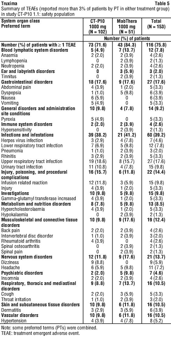
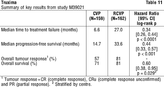
Results from three other randomised studies using rituximab in combination with chemotherapy regimens other than CVP (CHOP, MCP, CHVP/interferon-alfa 2a) have also demonstrated significant improvements in response rates, time dependent parameters as well as in overall survival (see Table 12).
Maintenance therapy.

Relapsed/refractory follicular NHL.

In a prospective, open label, international, multicentre, Phase III trial, 465 patients with relapsed/refractory follicular NHL were randomised in a first step to induction therapy with either CHOP (cyclophosphamide, doxorubicin, vincristine, prednisolone; n = 231) or rituximab plus CHOP (R-CHOP, n = 234), one dose of rituximab combined with each cycle of chemotherapy. The two treatment groups were well balanced with regard to baseline characteristics and disease status. A total of 334 patients achieving a complete or partial remission following induction therapy were randomised in a second step to rituximab maintenance therapy (n = 167) or observation (n = 167). Rituximab maintenance treatment consisted of a single infusion of rituximab at 375 mg/m2 body surface area given every 3 months until disease progression or for a maximum period of two years. Patients with hypogammaglobulinaemia (IgG < 3 g/L) or known HIV infection were excluded from the trial.
The final efficacy analysis included all patients randomised to both parts of the study. After a median observation time of 31 months for patients randomised to the induction phase, R-CHOP significantly improved the outcome of patients with relapsed/refractory follicular NHL when compared to CHOP (see Table 13).
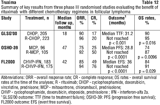
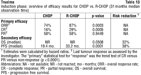
For patients randomised to the maintenance phase of the trial, the median observation time was 28 months from maintenance randomisation. Maintenance treatment with rituximab led to a clinically relevant and statistically significant improvement in the primary endpoint, PFS, (time from maintenance randomisation to relapse, disease progression or death) when compared to observation alone (p < 0.0001 log-rank test).
The median PFS was 42.2 months in the rituximab maintenance arm compared to 14.3 months in the observation arm. Using a cox regression analysis, the risk of experiencing progressive disease or death was reduced by 61% with rituximab maintenance treatment when compared to observation (95% CI; 45%-72%). Kaplan-Meier estimated progression-free rates at 12 months were 78% in the rituximab maintenance group vs. 57% in the observation group. An analysis of overall survival confirmed the significant benefit of rituximab maintenance over observation (p = 0.0039 log-rank test). Rituximab maintenance treatment reduced the risk of death by 56% (95% CI; 22%-75%).
The median time to new anti-lymphoma treatment was significantly longer with rituximab maintenance treatment than with observation (38.8 months vs. 20.1 months, p < 0.0001 log rank test). The risk of starting a new treatment was reduced by 50% (95% CI; 30%-64%). In patients achieving a CR/CRu (complete response unconfirmed) as best response during induction treatment, rituximab maintenance treatment significantly prolonged the median disease free survival (DFS) compared to the observation group (53.7 vs. 16.5 months, p = 0.0003) log-rank test (see Table 14). The risk of relapse in complete responders was reduced by 67% (95% CI; 39%-82%).
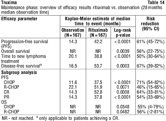
The benefit of rituximab maintenance treatment was confirmed in all subgroups analysed, regardless of induction regimen (CHOP or R-CHOP) or quality of response to induction treatment (CR or PR). Rituximab maintenance treatment significantly prolonged median PFS in patients responding to CHOP induction therapy (median PFS 37.5 months vs.11.6 months, p < 0.0001) as well as in those responding to R-CHOP induction (median PFS 51.9 months vs. 22.1 months, p = 0.0071). Although analysed subgroups were small, and the median survival had not been reached after an overall median observation period of 47.2 months, a clinically meaningful benefit in terms of overall survival was observed for patients receiving rituximab maintenance treatment when compared to observation, in the overall population.
Rituximab maintenance treatment provided consistent benefit in all subgroups tested [gender (male, female), age (≤ 60 years, > 60 years), stage (III, IV), WHO performance status (0 versus > 0), B symptoms (absent, present), bone marrow involvement (no versus yes), IPI (0-2 versus 3-5), FLIPI score (0-1, versus 2 versus 3-5), number of extra-nodal sites (0-1 versus > 1), number of nodal sites (< 5 versus ≥ 5), number of previous regimens (1 versus 2), best response to prior therapy (CR/PR versus NC/PD), haemoglobin (< 12 g/dL versus ≥ 12 g/dL), β2 microglobulin (< 3 mg/L versus ≥ 3 mg/L), LDH (elevated, not elevated) except for the small subgroup of patients with bulky disease.

Previously untreated follicular NHL.

In a prospective, open label, international, multi-centre, Phase III trial, 1193 patients with previously untreated advanced follicular lymphoma received induction therapy with R-CHOP (n = 881), R-CVP (n = 268) or R-FCM (n = 44), according to the investigators' choice. A total of 1078 patients responded to induction therapy, of which 1018 were randomised to rituximab maintenance therapy (n = 505) or observation (n = 513). The two treatment groups were well balanced with regards to baseline characteristics and disease status. Rituximab maintenance treatment consisted of a single infusion of rituximab at 375 mg/m2 body surface area given every 2 months until disease progression or for a maximum period of two years.
After a median observation time of 25 months from randomisation, maintenance therapy with rituximab resulted in a clinically relevant and statistically significant improvement in the primary endpoint of investigator assessed progression-free survival (PFS) as compared to no maintenance therapy in patients with previously untreated follicular NHL (see Table 15). This improvement in PFS was confirmed by an independent review committee (IRC) (see Table 15).
Significant benefit from maintenance treatment with rituximab was also seen for the secondary endpoints event-free survival (EFS), time to next anti-lymphoma treatment (TNLT), time to next chemotherapy (TNCT) and overall response rate (ORR) (see Table 15). Based on the limited number of deaths (58/513 patients (11%) in the observation arm and 59/505 patients (12%) in the rituximab maintenance arm), the current analysis did not show an advantage of maintenance treatment with rituximab in terms of overall survival (OS) HR 1.02 (95% CI: 0.71‐1.47; p = 0.8959).
The updated analysis corresponding to a median observation time of 73 months from randomisation confirm the results of the primary analysis (see Table 15).
Rituximab maintenance treatment provided consistent benefit in all subgroups tested: gender (male, female), age (< 60 years, ≥ 60 years), FLIPI score (1, 2 or 3), induction therapy (R-CHOP, R-CVP or R-FCM) and regardless of the quality of response to induction treatment (CR or PR).
There are currently no data to support superior efficacy for maintenance treatment given every 2 months over maintenance therapy given every 3 months, in either the relapsed/refractory or previously untreated setting.

Diffuse large B-cell non-Hodgkin's lymphoma.

In a randomised, Phase III, open-label trial, a total of 399 previously untreated elderly ambulatory patients (age 60 to 80 years, ECOG performance status 0-2) with moderate to advanced (Ann Arbor stage II-IV) diffuse large B-cell lymphoma received standard CHOP chemotherapy (cyclophosphamide 750 mg/m2, doxorubicin 50 mg/m2, vincristine 1.4 mg/m2 up to a maximum of 2 mg on day 1, and prednisone 40 mg/m2/day on days 1-5) every 3 weeks for eight cycles, or rituximab 375 mg/m2 administered as an intravenous infusion plus CHOP (R-CHOP). Rituximab was administered on the first day of the treatment cycle.
The final efficacy analysis included all randomised patients (197 CHOP, 202 R-CHOP), and had a median follow-up duration of approximately 31 months. The two treatment groups were well balanced in baseline characteristics and disease status. The final analysis confirmed that R-CHOP significantly increased the duration of event-free survival (the primary efficacy parameter, where events were death, relapse or progression of lymphoma, or institution of a new anti-lymphoma treatment) (p = 0.0001). Kaplan Meier estimates of the median duration of event-free survival were 35 months in the R-CHOP arm compared to 13 months in the CHOP arm, representing a risk reduction of 41%. At 24 months, estimates for overall survival were 68.2% in the R-CHOP arm compared to 57.4% in the CHOP arm. A subsequent analysis of the duration of overall survival, carried out with a median follow-up duration of 38 months, confirmed the benefit of R-CHOP over CHOP treatment (p = 0.0094), representing a risk reduction of 33%.
The analysis of all secondary parameters (response rates, progression-free survival, disease-free survival, duration of response) verified the treatment effect of R-CHOP compared to CHOP. The complete response rate after cycle 8 was 76.2% in the R-CHOP group and 62.4% in the CHOP group (p = 0.0028). The risk of disease progression was reduced by 46% and the risk of relapse by 51%.
In all patient subgroups (gender, age, age-adjusted IPI, Ann Arbor stage, ECOG, Beta 2 Microglobulin, LDH, Albumin, B-symptoms, Bulky disease, extranodal sites, bone marrow involvement), the risk ratios for event-free survival and overall survival (R-CHOP compared with CHOP) were less than 0.83 and 0.95 respectively, although the benefit with R-CHOP was not always statistically significant.
A subsequent analysis of the duration of overall survival, carried out with a median follow-up duration of 60 months, confirmed the benefit of R-CHOP over CHOP treatment (p = 0.0071), representing a risk reduction of 32%.

Chronic lymphocytic leukaemia (CLL).

In two open-label randomised studies, a total of 817 previously untreated patients and 552 patients with relapsed/refractory CLL were randomised to receive either fludarabine and cyclophosphamide (FC) chemotherapy (fludarabine 25 mg/m2, cyclophosphamide 250 mg/m2, days 1-3) every 4 weeks for 6 cycles or rituximab in combination with FC (R-FC). Rituximab was administered at a dosage of 375 mg/m2 during the first cycle one day prior to chemotherapy and at a dosage of 500 mg/m2 on day 1 of cycles 2-6. A total of 810 patients (403 R-FC, 407 FC) from the first-line study (see Table 16) and 552 patients (276 R-FC, 276 FC) for the relapsed/refractory study (see Table 18) were analysed for efficacy.
In the first-line study, after a median observation time of 20.7 months, the primary endpoint of progression-free survival (PFS) was a median of 40 months in the R-FC group and a median of 32 months in the FC group (p < 0.0001, log-rank test). The analysis of overall survival demonstrated improved survival in favour of the R-FC arm (p = 0.0427). These results were confirmed with longer follow-up: after a median observation time of 48.1 months, the median PFS was 55 months in the R-FC group and 33 months in the FC group (p < 0.0001, log-rank test) and overall survival analyses continued to show a significant benefit of R-FC treatment over FC chemotherapy alone (p = 0.0319, log-rank test). The benefit in terms of PFS was consistently observed in most patient subgroups analysed according to disease risk at baseline (i.e. Binet stages A-C) and was confirmed with longer follow-up (see Table 17).
In a case series of 30 previously untreated patients with CLL, an overall response rate of 97% was achieved with rituximab in combination with fludarabine, cyclophosphamide and mitoxantrone (FCM). Survival was not reported. In another case series of 64 previously untreated patients with CLL, an overall response rate of 91% and a median PFS of 32.6 months were achieved with rituximab in combination with pentostatin and cyclophosphamide (PC).
In the relapsed/refractory study, the median PFS (primary endpoint) was 30.6 months in the R-FC group and 20.6 months in the FC group (p = 0.0002, log-rank test). The benefit in terms of PFS was observed in almost all patient subgroups analysed according to disease risk at baseline. A non-significant trend towards improvement in overall survival was reported in the R-FC arm compared to the FC arm.
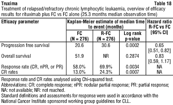
In relapsed/refractory CLL patients, response rates of 70% or greater have been reported in small studies of the following chemotherapy regimens with rituximab: FCM (fludarabine, cyclophosphamide, mitoxantrone), PC (pentostatin, cyclophosphamide), PCM (pentostatin, cyclophosphamide, mitoxantrone), CHOP (cyclophosphamide, doxorubicin, vincristine, prednisolone), bendamustine and cladribine.

Rheumatoid arthritis.

The efficacy and safety of rituximab in alleviating the symptoms and signs of RA was demonstrated in three randomised, controlled, double-blind, multicentre studies.
Study 1, WA17042 (REFLEX), was a double blind comparative study which included 517 patients that had experienced an inadequate response or intolerance to one or more TNF inhibitor therapies. Eligible patients had severe active RA, diagnosed according to the criteria of the American College of Rheumatology (ACR). The study population was comprised of adult patients aged ≥ 18 years with RA for at least 6 months who had experienced an inadequate response to previous treatment with an anti-TNF therapy. The primary endpoint was the percent of patients who achieved an ACR20 response at week 24. Patients received 2 x 1000 mg IV infusions of rituximab, each following an IV infusion of 100 mg methylprednisone and separated by an interval of 15 days. All patients received concomitant oral methotrexate (MTX) (10-25 mg/week) and 60 mg oral prednisone on days 2-7 and 30 mg on days 8-14 following the first infusion. Patients were followed beyond week 24 for long term endpoints, including radiographic assessment at 56 weeks. During this time patients could receive further courses of rituximab under an open label extension study protocol (see Radiographic response below).
Study 2, WA17043 (DANCER), was a randomised, double-blind, double-dummy, controlled, 3 x 3 multifactorial study which compared two different dose levels of rituximab (2 x 1000 mg or 2 x 500 mg) given with or without one of two corticosteroid infusion regimens in combination with weekly MTX. All patients received concomitant oral methotrexate. The primary endpoint was the proportion of RF (Rheumatoid Factor) positive patients with an ACR20 response at week 24. The study population was comprised of adult patients aged ≥ 18 years with RA who had previously failed 1-5 DMARDs and who currently had an inadequate response to MTX.
Study 3 was a double-blind, double-dummy, controlled study evaluating rituximab monotherapy, and rituximab in combination with either cyclophosphamide or MTX in patients with active RA who had not responded to one or more prior DMARDs. The primary endpoint was the proportion of patients with an ACR50 response at week 24. The study population was comprised of adult patients aged ≥ 21 years with RA who had failed 1-5 DMARDs, were RF seropositive at screening, and who currently had a partial clinical response to MTX monotherapy.
An ACR20 response was defined as at least a 20% improvement, compared to baseline, in both swollen and tender joint counts (SJC and TJC), as well as in 3 out of 5 additional parameters: physician's global assessment of disease activity, patient's global assessment of disease activity, patient's assessment of pain, Health Assessment Questionnaire Disability Index (HAQ-DI) and C-reactive protein (CRP).
The comparator drug in all three studies was weekly MTX (10-25 mg weekly).

Disease activity outcomes.

In all three studies, rituximab 2 x 1000 mg + MTX significantly increased the proportion of patients achieving at least a 20% improvement in ACR score compared with patients treated with MTX alone (see Table 19). The treatment effect was similar in patients independent of age, gender, body surface area, race, number of prior treatments or disease status.
Clinically and statistically significant improvement was also noted on all individual components of the ACR response (tender and swollen joint counts, patient and physician global assessment, disability index scores (HAQ), pain assessment and CRP (mg/dL).
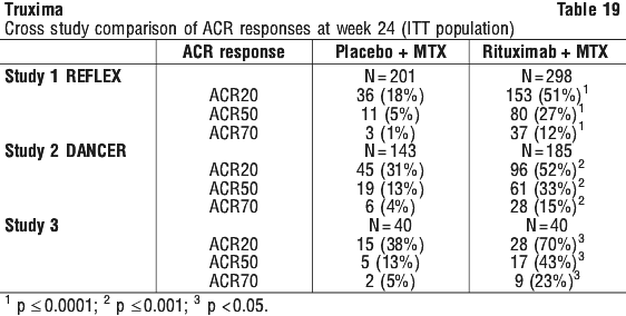
Rituximab + MTX treated patients had a significantly greater reduction in disease activity score (DAS28) than patients treated with MTX alone. A good to moderate EULAR response was achieved by significantly more rituximab + MTX treated patients compared to patients treated with MTX alone (see Table 20).

Radiographic response.

In Study WA17042 (REFLEX), structural joint damage was assessed radiographically and expressed as changes in Genant-modified Total Sharp Score (TSS) and its components, the erosion score (ES) and the joint space narrowing (JSN) score. Rituximab + MTX slowed the progression of structural damage compared to placebo + MTX after 1 year (see Table 24). 70% of patients initially randomised to rituximab + MTX and 72% of patients initially randomised to placebo + MTX were evaluated radiographically at year 2. Progression of structural damage in rituximab + MTX patients was further reduced in the second year of treatment (see Table 21).
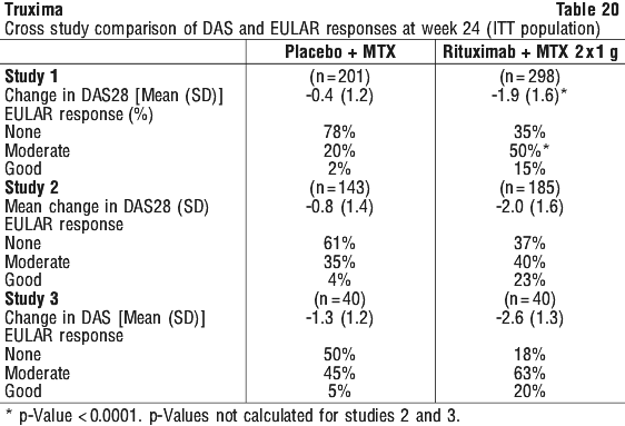
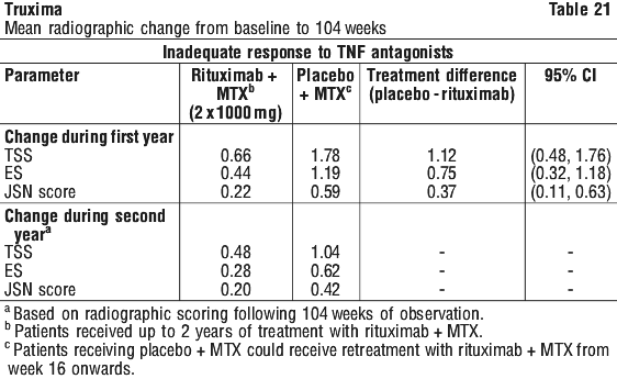
Following 2 years of treatment with rituximab + MTX, 57% of patients had no progression of structural damage. During the first year, 60% of rituximab + MTX treated patients had no progression, defined as a change in TSS of zero or less compared to baseline, compared to 46% of placebo + MTX treated patients. In their second year of treatment with rituximab + MTX, more patients had no progression than in the first year (68% vs. 60%), and 87% of the rituximab + MTX treated patients who had no progression in the first year also had no progression in the second year.

Quality of life outcomes.

Rituximab + MTX treated patients reported an improvement in all patient-reported outcomes such as Health Assessment Questionnaire Disability Index (HAQ-DI), Functional Assessment of Chronic Illness Therapy-Fatigue (FACIT-F) and Short Form-36 (SF-36) questionnaires. Significant reductions in disability index (HAQ-DI), fatigue (FACIT-F) (see Table 22), and improvement in both the physical health score (PHS) and mental health score (MHS) of the SF-36 were observed in patients treated with rituximab + MTX compared to patients treated with MTX alone.
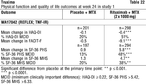
At week 24, in all three studies, the proportion of rituximab + MTX treated patients showing a clinically relevant improvement in HAQ-DI (defined as an individual total score decrease of > 0.25) was higher than among patients receiving MTX alone.

Laboratory evaluations.

Approximately 10% of patients with RA tested positive for HACA (Human Anti-Chimeric Antibody) in clinical studies. The emergence of HACA was not associated with clinical deterioration or with an increased risk of reactions to subsequent infusions in the majority of patients. The presence of HACA may be associated with worsening of infusion or allergic reactions after the second infusion of subsequent courses, and failure to deplete B cells after receipt of further treatment courses has been observed rarely.
In Study 1 WA17042 (REFLEX), 15/308 (4.8%) rituximab + MTX treated patients and 8/209 (3.8%) patients treated with MTX alone were anti-nuclear antibody (ANA) negative at day 1 and became ANA positive at week 16 and/or week 24. The adverse event profile in these patients did not provide any evidence of new onset autoimmune disease.
In RF positive patients, marked decreases were observed in RF concentrations following treatment with rituximab in all three studies (range 45-64%).
Hyperuricaemia (Grade 3/4) occurred in 143/950 (15%) patients, with the majority post-infusion on days 1 and/or 15. It was not associated with any clinical symptoms, and none of these patients developed evidence of renal disease. Increases in serum uric acid are often associated with the catabolism of DNA. This finding is consistent with the destruction of B cells resulting from rituximab therapy.
Hypophosphataemia (Grade 3) occurred in 193/950 (21%) patients. There was also one case of Grade 4 hypophosphataemia. Most cases occurred post-infusion, where patients received oral and/or IV corticosteroids. Low phosphate levels are associated with corticosteroid treatment and osteoporosis.
Plasma total immunoglobulin concentrations, total lymphocytes counts, and white cells generally remained within normal limits following rituximab treatment, with the exception of a transient drop in white cell counts over the first four weeks following therapy. Lymphopenia (Grade 3/4) was experienced by 679/1003 (68%) of patients compared to 52%-54% of patients who experienced Grade 3 lymphopenia and 1%-3% of patients who experienced Grade 4 lymphopenia in the 24-week double-blind populations. Most cases occurred immediately after the first infusion, consistent with peripheral B-cell depletion, and lymphocyte numbers recovered thereafter. The majority of the Grade 4 cases were transient, though 6 patients had more persistent Grade 4 lymphopenia, one of whom had a serious infection (2 occurrences of pneumonia in a diabetic patient; both cases resolved). All 6 patients had low lymphocyte counts before exposure to rituximab, including 2 patients who experienced up to Grade 4 lymphopenia whilst on placebo. A total of 17 non serious infections were reported, all of which resolved without sequelae. Neutropenia (Grade 3/4) was also observed at a higher rate in rituximab-treated patients compared to placebo (0.94% vs. 0.27%) (see Section 4.8 Adverse Effects (Undesirable Effects), Experience from clinical trials in rheumatoid arthritis, Laboratory abnormalities).
Titres of IgG antigen specific antibody to mumps, rubella, varicella, tetanus toxoid, influenza and streptococcus pneumococci remained stable over 24 weeks following exposure to rituximab in RA patients.
The effect of rituximab on a variety of biomarkers was evaluated in patients enrolled into Study 3. This substudy evaluated the impact of a single treatment course of rituximab on levels of biochemical markers, including markers of inflammation [Interleukin 6, C Reactive protein, Serum amyloid type A protein, Protein S100 isotypes A8 and A9], autoantibody (RF and anti-CCP immunoglobulin) production and bone turnover [osteocalcin and procollagen 1 N terminal peptide (P1NP)]. Rituximab treatment, whether as monotherapy or in combination with MTX or cyclophosphamide, reduced the levels of inflammatory markers significantly, relative to MTX alone, over the first 24 weeks of follow-up. Levels of markers of bone turnover, osteocalcin and P1NP, increased significantly in the rituximab + MTX groups compared to MTX alone.

Multiple course therapy.

Following completion of the 24-week double blind comparative study period, patients were permitted to enrol into an open-label long term follow up study. Patients received subsequent courses of rituximab as needed according to the treating clinician's assessment of disease activity and irrespective of the peripheral B lymphocyte count.
The all exposure population in the three double blind controlled trials (one Phase III and two Phase II trials) was 990 patients. Of these, 301 patients received a second course of rituximab 2 x 1000 mg + MTX, and 46 patients received a third course of rituximab 2 x 1000 mg + MTX.
At the point of data cut-off, 24.7% (193/781) of patients who had enrolled in the rituximab 2 x 1000 mg + MTX arms of the Phase II and Phase III studies had been retreated (point of data cut-off was defined as the time when all patients had been followed up for at least 24 weeks). Also at the data cut-off point, the majority of patients from the double blind comparative study period had received one course of treatment in the year. Kaplan-Meier analysis of time to second treatment course (censoring patients who did not receive a second treatment course or who withdrew from the study) shows an estimated median time for retreatment in the prior anti-TNF population of 364 days (interquartile range: 245-559 days), Figure 1, and 547 days (interquartile range: 302-889 days) in the no prior anti-TNF population, Figure 2.
The time interval between courses was variable. The majority of patients, who had two treatment courses at the time of cut-off, received their second course of treatment 6 to 12 months after the first treatment course. Some patients required even less frequent retreatment. The response to further therapy was at least the same magnitude as that following the initial treatment course, as evidenced by the change from baseline DAS28 (Figure 3).
Since many patients in the prior anti-TNF population remain in the studies after a single course of treatment with rituximab + MTX, these results are subject to change as the observation period increases.

120 Minute infusion rate study (ML25641) in rheumatoid arthritis.

Study 4, a multi-centre, open-label single-arm trial, 351 patients with moderate to severe active RA, who had an inadequate response to at least one TNF inhibitor and were receiving MTX, were to receive 2 courses of rituximab treatment. Patients who were naïve to prior rituximab therapy (n = 306) and those who had received 1 to 2 prior courses of rituximab 6-9 months prior to baseline (n = 45) were eligible for enrollment.
Patients received 2 courses of rituximab 2 x 1000 mg + MTX treatment, the first course was administered on Days 1 and 15 and the second course 6 months later on Days 168 and 182. The first infusion of the first course (Day 1 infusion) was administered over a 4.25 hour period. The second infusion of the first course (Day 15 infusion) and both infusions in the second course (Day 168 and 182 infusions) were administered over 120 minutes. Any patient experiencing a serious infusion-related reaction with any infusion was withdrawn from the study. In this study, an infusion-related reaction (IRR) was defined as any adverse event that occurred during or within 24 hours following the infusion of rituximab and met pre-specified criteria for adverse event terms for IRRs. IRRs were defined as serious if they met one of the following seriousness criteria: fatal, life-threatening, required inpatient hospitalisation or prolongation of existing hospitalisation, resulted in persistent or significant disability/incapacity, were medically significant.
The primary objective of this study was to assess the safety of administering the second infusion of the first study course of rituximab over 120 minutes in rheumatoid arthritis patients.
The incidence of IRRs at Day 15 was 6.5% (95% CI [4.1%-9.7%]) consistent with the rate observed historically. There were no serious IRRs observed. Data observed for the infusions on Days 168 and 182 (120 minute infusion) demonstrates a low incidence of IRRs, similar to the rates observed historically, with no serious IRRs occurring (see Section 4.8 Adverse Effects (Undesirable Effects), Experience from clinical trials in rheumatoid arthritis).

Granulomatosis with polyangiitis (Wegener's) and microscopic polyangiitis.

A total of 197 patients with severely, active GPA and MPA were enrolled and treated in study ITN021AI (RAVE), a phase II/III, active controlled, randomised, double-blind, multicenter, non-inferiority study. Patients were 15 years of age or older, diagnosed with severely, active GPA (75% of patients) or MPA (24% of patients) according to the Chapel Hill Consensus conference criteria (1% of patients had unknown GPA and MPA type).
Patients were randomised in a 1:1 ratio to receive either oral cyclophosphamide (CYC) daily (2 mg/kg/day) for 3-6 months followed by azathioprine, or rituximab (375 mg/m2) once weekly for 4 weeks. Patients in both arms received 1000 mg of pulse IV methylprednisolone (or another equivalent-dose glucocorticoid) per day for 1 to 3 days, followed by oral prednisone (1 mg/kg/day, not exceeding 80 mg/day). Prednisone tapering was to be completed by 6 months from the start of study treatment. For prophylaxis against Pneumocystis jiroveci pneumonia, patients in both treatment arms received trimethoprim/sulfamethoxazole unless contraindicated.
Study exclusion criteria included diagnosis of Eosinophilic Granulomatosis with polyangiitis (Churg Strauss syndrome), active infection (including active or documented hepatitis B or C), severe hepatic impairment or significant renal impairment (serum creatinine > 4.0). Subjects requiring mechanical ventilation were also excluded.
Patient demographic characteristics were similar between the treatment arms in terms of age at disease onset, gender, primary race, and ethnicity. The median patient age was 52.0 years with a range of 15 to 92 years. At baseline disease assessment, the majority of patients in each arm had pulmonary, systemic, or ear/nose/throat involvement. 65/99 patients in the rituximab arm and 65/98 patients in the CYC arm had renal involvement. Other disease characteristics were generally balanced between the two treatment arms with the exception of estimated creatinine clearance. Mean (SD) creatinine clearance (estimated by Cockcroft-Gault formula) was 76.51 (46.27) and 91.40 (49.24) mL/min for the rituximab and CYC arms, respectively. The median values were also lower in the rituximab arm compared with the CYC arm (67.61 vs. 87.47 mL/min, respectively).
The primary outcome measure was achievement of complete remission (CR) at 6 months defined as a Birmingham Vasculitis Activity Score for Wegener's Granulomatosis (BVAS/WG) of 0, and off glucocorticoid therapy. The prespecified non-inferiority margin for the treatment difference was 20%. The study demonstrated non-inferiority of rituximab to CYC for CR at 6 months (see Table 23). In addition, the CR rate in the rituximab arm was significantly greater than the estimated CR rate in patients with severe GPA and MPA not treated or treated only with glucocorticoids, based on historical control data.
Efficacy was observed both for patients with newly diagnosed GPA and MPA and for patients with relapsing disease. As RAVE did not investigate alternative rituximab doses or infusion schedules, and no prior dose-finding studies were performed, an optimal dose has not been determined. Since corticosteroids were not used in all patients beyond 6 months, no conclusions can be drawn on the need for additional use. Any further corticosteroid use should be at the physician's discretion.

Retreatment.

Based upon investigator judgment, 15 patients received a second course of rituximab therapy for treatment of relapse of disease activity which occurred between 6 and 18 months after the first course of rituximab. The limited data from the present study preclude any conclusions regarding the efficacy of subsequent courses of rituximab in patients with GPA or MPA.

Comparability of Truxima with MabThera.

The clinical comparability between Truxima and MabThera was demonstrated in two randomized, controlled, parallel-group, double blind, multicentre studies and one open-label, single-arm maintenance study in RA patients; and two randomized, controlled, parallel-group, double blind, multicenter study in AFL patients and in LTBFL patients, respectively.

Rheumatoid arthritis.

Study CT-P10 1.1.

A phase 1, randomized (2:1), controlled, multicenter, 2-arm, parallel-group, double blind study to demonstrate similarity of PK between CT-P10 and MabThera in patients with active RA (up to week 24) and to evaluate long-term efficacy, PD, overall safety, and biomarker of CT-P10 compared to MabThera (up to week 72). The study population was comprised of adult patients ≥ 21 years with active RA who have had an inadequate response or were intolerant to TNF inhibitors.
CT-P10 and MabThera were co-administered with MTX between 10 to 25 mg/week, orally or parenterally (dose and route had to be maintained from beginning to end of study) and folic acid ≥ 5 mg/week, oral dose.
A total of 154 patients were randomly assigned to study drug (103 patients and 51 patients in the CT-P10 and MabThera groups, respectively). Of these, 153 (99.4%) patients initiated the 1st treatment course in Core Study Period (102 [99%] patients and 51 [100%] patients in the CT-P10 and MabThera groups, respectively).
The efficacy endpoints were similar between CT-P10 and MabThera groups.

Study CT-P10 1.3.

CT-P10 1.3 (Extension study to CT-P10 1.1) was an open-label, single-arm, maintenance study to demonstrate long-term efficacy and safety of CT-P10 up to 104 weeks in patients with RA who were treated with CT-P10 or MabThera in Study CT-P10 1.1. The study population was comprised of adult patients ≥ 21 years with RA who have completed the scheduled visits in Study CT-P10 1.1.
A total of 87 patients (58 [100%] in CT-P10 maintenance group and 29 [100%] in CT-P10 switch group) who had completed the main study CT-P10 1.1 were recruited into Study CT-P10 1.3 (ITT population). Of these, 38 (65.5%) patients who had received CT-P10 and 20 (69.0%) who had received MabThera in Study CT-P10 1.1 were treated with CT-P10 in the maintenance and switch groups, respectively. Among these 58 patients, a total of 22 patients (13 [34.2%] in CT-P10 maintenance group and 9 [45.0%] in CT-P10 switch group) received 1 treatment course in Study CT-P10 1.1 while a total of 36 patients (25 [65.8%] in the CT-P10 maintenance group and 11 [55.0%] in the CT-P10 switch group) received 2 treatment courses in Study CT-P10 1.1.
A total of 68 (78.2%) patients completed the study (47 [81.0%] in the CT-P10 maintenance group and 21 [72.4%] in the CT-P10 switch group). A similar proportion of patients in each treatment group discontinued the study (11 [19.0%] patients and 8 [27.6%] patients in the CT-P10 maintenance and switch groups, respectively).
The efficacy population consisted of 38 patients in the CT-P10 maintenance group and 19 patients in the CT-P10 switch group.
The efficacy endpoints were similar between CT-P10 and MabThera groups.

Study CT-P10 3.2.

A Phase 3, randomised (1:1:1), controlled, multicentre, 3-arm, parallel-group, double-blind, prospective study in patients with RA. The study population was comprised of adult patients ≥ 18 years with active RA who have had an inadequate response or were intolerant to TNF inhibitors.
This 3-way PK and efficacy study demonstrated PK equivalence between CT-P10, MabThera and Rituxan (Part 1) and efficacy equivalence between CT-P10 and combined reference products groups (MabThera/Rituxan) (Part 2) in patients with RA who received concomitant MTX and folic acid.
A dose of 1,000 mg of CT-P10, MabThera or Rituxan (IV) was co-administered with MTX given at a dose between 7.5 to 25 mg orally or parenterally every week (dose and route had to be maintained from the beginning to the end of the study) and folic acid at a dose of ≥ 5 mg/week. Patients were able to receive up to 3 treatment courses (2 courses in the Main Study Period and 1 additional course in the Extension Study Period). Each course consists of 2 infusions separated by a 2-week interval. In the third treatment course (1 additional course in the Extension Study Period), patients who received Rituxan in the Main Study Period will be re-randomised to either the Rituxan or CT-P10 treatment groups and patients who received MabThera in the Main Study Period will be switched to CT-P10 while patients who received CT-P10 will remain in CT-P10 group.
In Part 1, a total of 189 patients were randomly assigned to study drugs (64 patients in CT-P10 group, 60 in MabThera and 65 in Rituxan groups). All-randomised patients initiated treatments, and a total of 177/189 (93.7%) patients completed the 1st treatment course and a total of 170/189 (89.9%) completed the 2nd treatment course. A similar proportion of patients in each treatment group discontinued the study both after the 1st and 2nd treatment courses in Part 1.
In Part 2, a total of 372 patients were randomised to study drugs and initiated the treatments (161 patients in CT-P10 group, 60 patients in MabThera group and 151 patients in Rituxan group). Of these, 345/372 (92.7%) patients completed the 1st treatment course and a total of 330/372 (88.7%) completed the 2nd treatment course. A similar proportion of patients in each treatment group discontinued the study both after the 1st and 2nd treatment courses in Part 2.
The primary efficacy endpoint was defined as the change from baseline in disease activity measured by DAS28 (CRP) at Week 24 between the 2 treatment groups, CT-P10 and reference products groups.
In the efficacy population, the 95% CIs for the estimate of treatment difference was well within the pre-defined equivalence margin of 0.6 and hence, the therapeutic equivalence between CT-P10 and reference products group in terms of change from baseline in DAS28 (CRP) at Week 24 has been established at the 5% level of significance. A similar result was found in change from baseline of DAS28 (ESR), showing no significant difference between CT-P10 and reference products groups. Also, the same analysis has been carried out in the all-randomised population as a sensitivity analysis and the results were in line with that of efficacy population (see Table 25).

Advanced follicular lymphoma.

Study CT-P10 3.3.

Study CT-P10 3.3 was a randomized, double-blind, parallel-group, active-controlled, comparative study in which patients with AFL were randomized in a 1:1 ratio to receive either CT-P10 or Rituxan in combination with CVP (cyclophosphamide, vincristine, and prednisolone) chemotherapy. CT-P10 or Rituxan (375 mg/m2 IV) was administered in combination with CVP (cyclophosphamide 750 mg/m2 IV, vincristine 1.4 mg/m2 [maximum 2 mg] IV, and prednisolone 40 mg/m2 PO) once every 3 weeks during Core Study Period (up to 8 cycles). Subsequently, CTP10 or Rituxan was administered alone every 2 months during maintenance study period (up to 2 years).
A total number of 140 patients including 121 patients from Part 1 were randomised to receive either CT-P10 (N = 70) or Rituxan (N = 70). All randomised patients in the CT-P10 and Rituxan treatment groups initiated study treatment and the majority of patients completed the Core Study Period (62 [88.6%] patients in each group) except 16 patients (8 [11.4%] patients in each group, respectively) who discontinued before completion of the Core Study Period. The most frequently reported reasons for discontinuation from the Core Study Period were progressive disease (2 [2.9%] patients and 3 [4.3%] patients in the CT-P10 and Rituxan groups, respectively) and adverse event (4 [5.7%] patients and 1 [1.4%] patient in the CT-P10 and Rituxan groups, respectively).
The primary efficacy endpoint for Study CT-P10 3.3 assessed in Part 2 of the study, ORR (CR + CRu + PR) according to 1999 IWG criteria over Cycle 8 in PP population is presented in Table 26. The proportions of patients achieving ORR according to 1999 IWG criteria were 97.0% (64/66 patients) and 92.6% (63/68 patients) in the CT P10 and Rituxan groups, respectively.
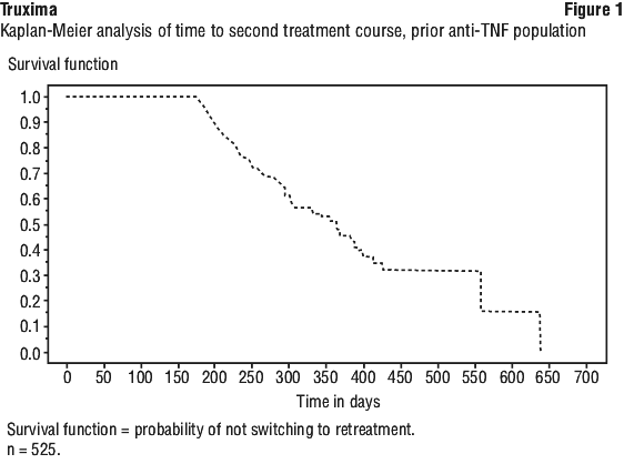
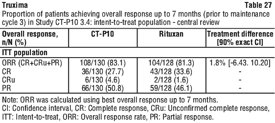
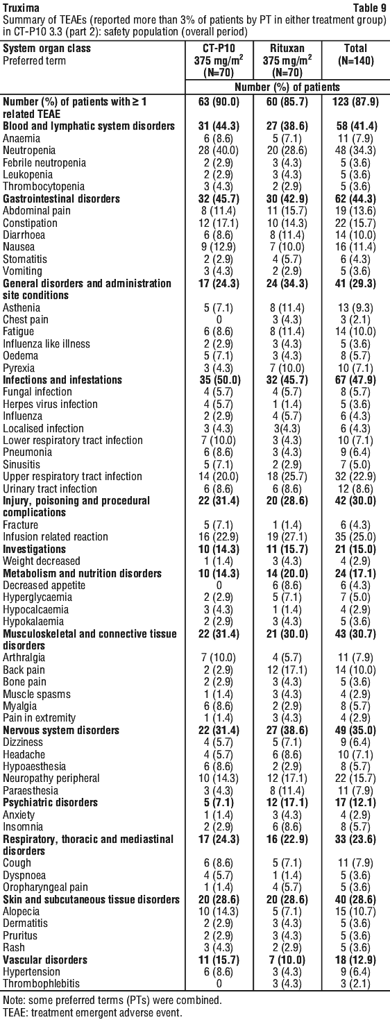
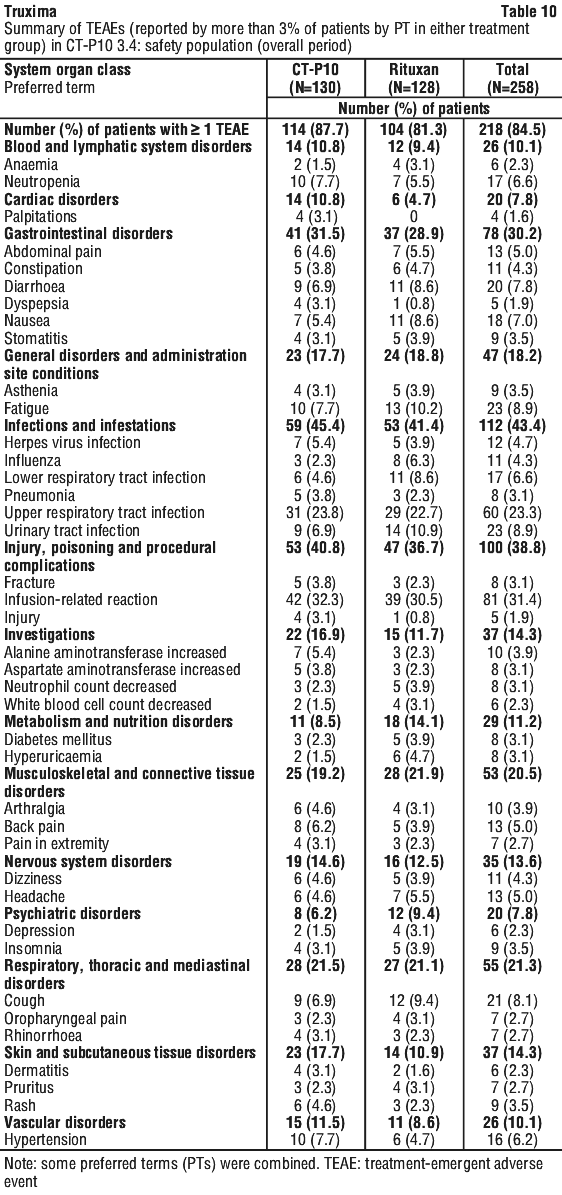
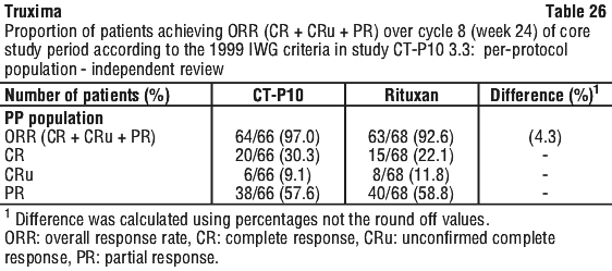
In Study CT-P10 3.3, efficacy in terms of overall response rate using the 1999 IWG criteria was evaluated over a 24 week period and efficacy was comparable between the CT-P10 and Rituxan groups.

Low tumor burden follicular lymphoma.

Study CT-P10 3.4.

Study CT-P10 3.4 was a randomized, double-blind, parallel-group, active-controlled, comparative study in which patients with LTBFL were randomized in a 1:1 ratio to receive either CT-P10 or Rituxan as monotherapy. All subjects received either CT-P10 or Rituxan (375 mg/m2 IV) once weekly for 4 weeks during the induction cycle and every 8 weeks for 6 cycles during the maintenance cycle. Subsequently, all patients were allowed to receive CT-P10 every 8 weeks for 6 additional cycles.
A total number of 258 patients were randomised to receive either CT-P10 (N=130) or Rituxan (N=128) and initiated Induction Study Period. The majority of patients (128 [98.5%] for CT-P10 and 128 [100.0%] for Rituxan) completed Induction Study Period and most of the patients (123 [94.6%] for CT-P10 and 120 [93.8%] for Rituxan) initiated Maintenance Study Period. Up to 1st year of the Maintenance Study Period, the most frequently reported reason for discontinued study treatment was progressive disease (6 [4.6%] for CT-P10 and 12 [9.4%] for Rituxan).
This study used an equivalence design for the primary efficacy endpoint, ORR (CR + CRu + PR) at 7 months (prior to maintenance cycle 3) according to the Modified Response Criteria for Malignant Lymphoma, with the pre-defined equivalence margin. The difference in ORR from central review between CT-P10 and Rituxan was 1.8%. The 90% exact binomial CI for the difference in ORR between CT-P10 and Rituxan was within the pre-defined equivalence margin corresponding to a 5% significance level. This result strongly supported the therapeutic equivalence between CT-P10 and Rituxan from the primary analysis result. See Table 27.

5.2 Pharmacokinetic Properties

Non-Hodgkin's lymphoma.

Based on a population pharmacokinetic analysis in 298 NHL patients who received single or multiple infusions of rituximab as a single agent or in combination with CHOP therapy, the typical population estimates of nonspecific clearance (CL1), specific clearance (CL2) likely contributed by B cells or tumour burden, and central compartment volume of distribution (V1) were 0.14 L/day, 0.59 L/day, and 2.7 L, respectively. The estimated median terminal elimination half-life of rituximab was 22 days (range, 6.1 to 52 days). Baseline CD19-positive cell counts and size of measurable tumour lesions contributed to some of the variability in CL2 of rituximab in data from 161 patients given 375 mg/m2 as an intravenous (IV) infusion for 4 weekly doses. Patients with higher CD19-positive cell counts or tumour lesions had a higher CL2. However, a large component of inter-individual variability remained for CL2 after correction for CD19-positive cell counts and tumour lesion size. V1 varied by body surface area (BSA) and CHOP therapy. The variability in V1 caused by the range in BSA (1.53 to 2.32 m2) and concurrent CHOP therapy was relatively small (27.1% and 19%, respectively). Age, gender, race, and WHO (World Health Organisation) performance status had no effect on the pharmacokinetics of rituximab. This analysis suggests that dose adjustment of rituximab with any of the tested covariates is not expected to result in a meaningful reduction in its pharmacokinetic variability.
Rituximab at a dose of 375 mg/m2 was administered as an IV infusion at weekly intervals for 4 doses to 203 patients with NHL naive to rituximab. The mean Cmax following the fourth infusion was 486 microgram/mL (range 77.5 - 996.6 microgram/mL). The peak and trough serum levels of rituximab were inversely correlated with baseline values for the number of circulating CD19-positive B-cells and measures of disease burden. Median steady-state serum levels were higher for responders compared with non-responders. Serum levels were higher in patients with International Working Formulation (IWF) subtypes B, C, and D as compared with those with subtype A.
Rituximab was detectable in the serum of patients 3 - 6 months after completion of last treatment.
Rituximab at a dose of 375 mg/m2 was administered as an IV infusion at weekly intervals for 8 doses to 37 patients with NHL. The mean Cmax increased with each successive infusion, spanning from a mean of 243 microgram/mL (range, 16 - 582 microgram/mL) after the first infusion to 550 microgram/mL (range 171 - 1177 microgram/mL) after the eighth infusion.
The pharmacokinetic profile of rituximab when administered as 6 infusions of 375 mg/m2 in combination with 6 cycles of CHOP chemotherapy was similar to that seen with rituximab alone.

Chronic lymphocytic leukaemia (CLL).

Rituximab was administered as an IV infusion at a first-cycle dose of 375 mg/m2 increased to 500 mg/m2 each cycle for a further 5 doses in combination with fludarabine and cyclophosphamide (FC) in CLL patients. The mean Cmax (N = 15) was 408 microgram/mL (range, 97 - 764 microgram/mL) after the fifth 500 mg/m2 infusion.

Rheumatoid arthritis.

Following two intravenous infusions of rituximab at a dose of 1000 mg, two weeks apart, the mean terminal half-life was 20.8 days (range 8.58 to 35.9 days), mean systemic clearance was 0.23 L/day (range 0.091 to 0.67 L/day), and mean steady-state distribution volume was 4.6 L (range 1.7 to 7.51 L). Population pharmacokinetic analysis of the same data gave similar mean values for systemic clearance and half-life, 0.26 L/day and 20.4 days, respectively. Population pharmacokinetic analysis revealed that BSA and gender were the most significant covariates to explain inter-individual variability in pharmacokinetic parameters. After adjusting for BSA, male subjects had a larger volume of distribution and a faster clearance than female subjects. The gender-related pharmacokinetic differences are not considered to be clinically relevant and dose adjustment is not required.
The pharmacokinetics of rituximab were assessed following two IV doses of 500 mg and 1000 mg on days 1 and 15 in four studies. In all these studies, rituximab pharmacokinetics were dose proportional over the limited dose range studied. Mean Cmax for serum rituximab following first infusion ranged from 157 to 171 microgram/mL for 2 x 500 mg dose and ranged from 298 to 341 microgram/mL for 2 x 1000 mg dose. Following second infusion, mean Cmax ranged from 183 to 198 microgram/mL for the 2 x 500 mg dose and ranged from 355 to 404 microgram/mL for the 2 x 1000 mg dose. Mean terminal elimination half-life ranged from 15 to 16.5 days for the 2 x 500 mg dose group and 17 to 21 days for the 2 x 1000 mg dose group. Mean Cmax was 16 to 19% higher following second infusion compared to the first infusion for both doses.
Upon re-treatment with a second course, the pharmacokinetics of rituximab were again assessed following two IV doses of 500 mg and 1000 mg. Mean Cmax for serum rituximab following first infusion was 170 to 175 microgram/mL for 2 x 500 mg dose and 317 to 370 microgram/mL for 2 x 1000 mg dose. Cmax following second infusion, was 207 microgram/mL for the 2 x 500 mg dose and ranged from 377 to 386 microgram/mL for the 2 x 1000 mg dose. Mean terminal elimination half-life after the second infusion, following the second course, was 19 days for 2 x 500 mg dose and ranged from 21 to 22 days for the 2 x 1000 mg dose. PK parameters for rituximab were comparable over the two treatment courses.

Granulomatosis with polyangiitis (Wegener's) (GPA) and microscopic polyangiitis (MPA).

Based on the population pharmacokinetic analysis of data in 97 GPA and MPA patients who received 375 mg/m2 rituximab once weekly for four doses, the estimated median terminal elimination half-life was 23 days (range 9 to 49 days). Rituximab mean clearance and volume of distribution were 0.313 L/day (range 0.116 to 0.726 L/day) and 4.50 L (range 2.25 to 7.39 L), respectively. The PK parameters of rituximab in GPA and MPA patients appear similar to what has been observed in RA patients (see Section 5.2 Pharmacokinetic Properties, Rheumatoid arthritis).

Comparability of Truxima with MabThera.

PK equivalence between Truxima (CT-P10) and MabThera was demonstrated in 1 Phase 1 study and 2 Phase 3 studies (Studies CT-P10 1.1, CT-P10 3.2 in RA patients and CT-P10 3.3 in AFL patients) in the CT-P10 clinical development program.

Study CT-P10 1.1.

Study CT-P10 1.1 demonstrated PK equivalence in terms of area under the concentration-time curve from time zero to the last quantifiable concentration (AUC0-last [AUC covering both infusions, from time zero to the last measurable concentration at Week 24]) and the maximum serum concentration (Cmax) after the 2nd infusion between CT-P10 and MabThera in patients with RA.
The 90% CIs of ratios of geometric means in the PK population and geometric LS means in All PK population for both AUC0-last and Cmax were entirely contained in the equivalence range of 80% to 125% indicating that rituximab exposures from CT-P10 are similar to those from MabThera (see Table 28).

Study CT-P10 3.2.

Phase 3 study to compare the PK, efficacy and safety between CT-P10, Rituxan and MabThera in Patients with RA Study CT-P10 3.2 consisted of 2 Parts. Part 1 was designed for demonstration of 3-way PK equivalence between 1) CT-P10 and MabThera, 2) CT-P10 and Rituxan, 3) MabThera and Rituxan. Part 2 was intended to demonstrate therapeutic equivalence between CT-P10 and the combined reference products, MabThera and Rituxan.
In the PK population, the 90% CIs of ratios of geometric LS means for all primary PK endpoints (AUC0-last, AUC0-inf and Cmax) were entirely contained in the equivalence range of 80% to 125% indicating that rituximab exposures are similar between CT-P10, MabThera and Rituxan.

Study CT-P10 3.3.

Phase 3 study to demonstrate equivalence of pharmacokinetics and non-inferiority of efficacy for CT-P10 in comparison with Rituxan, each administered in combination with cyclophosphamide, vincristine and prednisone (CVP) in patients with AFL. Study CT-P10 3.3 has been designed to demonstrate PK similarity (Part 1) and therapeutic non-inferiority (Part 2) between CT-P10 and Rituxan in patients with AFL who received concomitant CVP.
In the PK population, the 90% CIs of ratios of geometric LS means for both AUCtau and Cmax,ss were entirely contained in the equivalence range of 80% to 125% indicating that rituximab exposures from CT-P10 are similar to those from Rituxan.

5.3 Preclinical Safety Data

Genotoxicity.

The genotoxic potential of rituximab has not been investigated.

Carcinogenicity.

The carcinogenic potential of rituximab has not been investigated.

6 Pharmaceutical Particulars

6.1 List of Excipients

The product is formulated in 7.35 mg/mL sodium citrate buffer containing 0.7 mg/mL polysorbate 80, 9.0 mg/mL sodium chloride and sterile water for injection. The pH is adjusted to 6.5 with sodium hydroxide or hydrochloric acid.

6.2 Incompatibilities

No incompatibilities between Truxima and polyvinyl chloride or polyethylene bags have been observed.

6.3 Shelf Life

In Australia, information on the shelf life can be found on the public summary of the Australian Register of Therapeutic Goods (ARTG). The expiry date can be found on the packaging.

6.4 Special Precautions for Storage

Truxima vials must be refrigerated between 2°C to 8°C. Do not freeze Truxima vials. Truxima vials must be protected from direct sunlight. Do not use beyond the expiry date stamped on the carton/vial. Truxima vials should be used once only and any unused portion left in the vials should be discarded.

6.5 Nature and Contents of Container

100 mg/10 mL.

Truxima 100 mg/10 mL is supplied as a concentrated solution for dilution and intravenous infusion in single-use type I glass vials with rubber stoppers and aluminium flip-off seal.
Truxima 100 mg/10 mL is available in packs of 2 vials.

500 mg/50 mL.

Truxima 500 mg/50 mL is supplied as a concentrated solution for dilution and intravenous infusion in single-use type I glass vials with rubber stoppers and aluminium flip-off seal.
Truxima 500 mg/50 mL is available in packs of 1 vial.

6.6 Special Precautions for Disposal

The release of medicines into the environment should be minimised. Medicines should not be disposed of via wastewater and disposal through household waste should be avoided. Unused or expired medicine should be returned to a pharmacy for disposal.

6.7 Physicochemical Properties

Truxima (rituximab) is a genetically engineered chimeric murine/human monoclonal antibody directed against the CD20 antigen found on the surface of normal and malignant B lymphocytes. The antibody is a glycosylated IgG1 kappa immunoglobulin containing murine light- and heavy-chain variable region sequences (Fab domain) and human constant region sequences (Fc domain). Rituximab is composed of 1,328 amino acids and has an approximate molecular weight of 144 kD. Rituximab has a high binding affinity for the CD20 antigen of 5.2 to 11.0 nanoM.
The chimeric anti-CD20 antibody is produced by mammalian (Chinese hamster ovary) cell suspension culture in a nutrient medium containing 100 mg/mL of the antibiotic gentamicin. The antibiotic is not detectable in the final product. The anti-CD20 antibody is purified by affinity chromatography and ion exchange, including specific viral inactivation and removal procedures.

CAS number.

174722-31-7.
Truxima is a biosimilar medicine to MabThera. The comparability of Truxima with MabThera has been demonstrated with regard to physicochemical characteristics and efficacy and safety outcomes [see Section 5 Pharmacological Properties, Clinical trials; Section 4.8 Adverse Effects (Undesirable Effects)]. The evidence for comparability supports the use of Truxima for the listed indications.

7 Medicine Schedule (Poisons Standard)

Prescription only medicine - Schedule 4.

Summary Table of Changes