Key points
- Requests for thyroid tests are increasing, although in many cases clinical need is not evident.
- If thyroid dysfunction is suspected, measuring TSH alone is recommended as the first step. Test T4 only when TSH is abnormal.
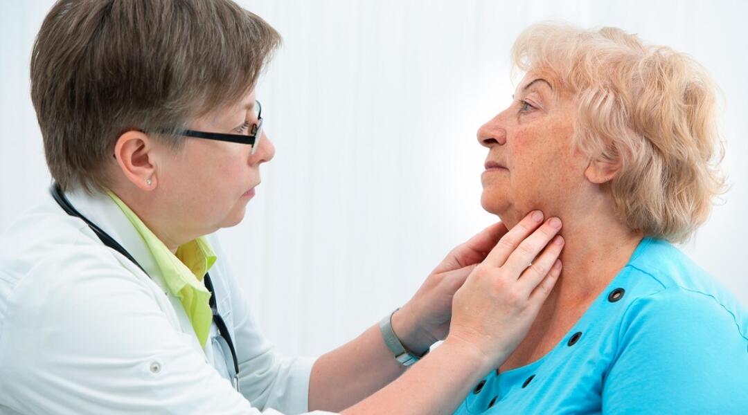
- Ultrasound should be considered for patients with thyroid dysfunction and a goitre (enlarged thyroid) or palpable thyroid nodules, but NOT if the only abnormality is hypothyroidism or elevated antithyroid antibodies.
- Repeat TSH testing within 12 months is generally not recommended if TSH is normal.
- Fatigue alone should not be the basis of thyroid testing. Instead, take a detailed clinical history and perform a targeted physical examination before requesting tests or treatment.
Thyroid problems in Australia
"Thyroid disorders are very common. As many symptoms of thyroid disease are non-specific, careful history taking and physical examination are key to avoiding unnecessary testing."
Associate Professor Shane Hamblin, Head of Endocrinology and Diabetes at Western Health, Victoria
The number of people undergoing thyroid testing in Australia is increasing at a faster rate than the population,2 and evidence indicates that at least some of these tests may be clinically unnecessary.3
Increased testing carries the risk of incidental findings which may lead to a cascade of tests and procedures, as well as uncertainty and anxiety for patients.3,4 To ensure the benefits of thyroid testing outweigh harms, adherence to testing guidelines is important.
Identifying thyroid disease in general practice
Thyroid disease can be broadly categorised as:
- thyroid dysfunction (hypothyroidism, hyperthyroidism,) or
- structural disease (goitre, nodules and cancer).
Hypothyroidism is the most common disorder of thyroid function, with a prevalence ranging from 0.5% to 5% for overt and subclinical cases, respectively.5 Hyperthyroidism (low TSH, high free T4/T3) is less common than hypothyroidism, with a prevalence of around 0.5%–1.0%.5-7
When to suspect thyroid dysfunction
Signs and symptoms of hypothyroidism may vary depending on patient characteristics and the severity of dysfunction. Classic symptoms include fatigue, weight gain, cold intolerance, arthralgia, constipation, menorrhagia, irregular menstrual cycles, and dry skin and hair.8 Physical signs include coarse skin and hair, bradycardia and goitre but these may be absent in mild hypothyroidism.8
Many of the signs and symptoms are not specific to hypothyroidism: around 20%–25% of people with normal TSH report one or two of these symptoms.9 Laboratory tests are required to establish a diagnosis of hypothyroidism.
Hypothyroidism can be classified as overt, subclinical or secondary (central).
- Overt hypothyroidism (high TSH, low free T4) is usually symptomatic and readily diagnosed, although there are exceptions.
- Subclinical hypothyroidism (elevated TSH, normal free T4) is more common than overt hypothyroidism, but less frequently detected clinically as many patients have vague symptoms.
- Secondary (or central) hypothyroidism is caused by disorders of the pituitary gland or hypothalamus and is characterised by decreased TSH and a disproportionately low concentration of free T4.10 Secondary hypothyroidism is uncommon, but in situations where a central cause is suspected (eg, known pituitary tumour or head injury) it is essential that T4 is checked as well as TSH, since TSH may be in the normal range.
Classic symptoms for hyperthyroidism include weight loss, heat intolerance, palpitations, breathlessness, anxiety, diarrhoea, menstrual disturbances, tremor and proximal muscle weakness. Physical signs include tremor, tachycardia, ophthalmopathy (if due to Graves disease), goitre and difficulty rising from a squatting position.8 However some patients (especially in older age groups), may present with ‘apathetic hyperthyroidism’ and lack many of the classical features.
- Overt hyperthyroidism is characterised by increased free T4 and free T3 and low TSH. It is most commonly caused by Graves disease, or toxic nodular goitre.
- Subclinical hyperthyroidism is characterised by suppressed TSH and normal free T3/T4 in patients with or without symptoms of hyperthyroidism.11,12 Mild subclinical hyperthyroidism (with low but detectable TSH levels) may be caused by autonomous thyroid nodules but may also be found in healthy individuals.8 Approximately 5% of individuals progress from subclinical to overt dysfunction yearly.13 Because of the diversity of clinical presentations of hyperthyroidism, laboratory testing is appropriate on a low index of suspicion.14
Current recommendations for monitoring, testing and treating thyroid dysfunction are summarised in Table 1.
Optimising the use of thyroid testing and imaging
Screening for thyroid dysfunction is not recommended in asymptomatic patients unless there is a clinical suspicion of thyroid dysfunction or the patient is in a high-risk group (such as patients with autoimmune disease or type 1 diabetes).15
Measuring TSH is recommended as the first-line test for possible thyroid dysfunction.1 If TSH is in the reference range, additional tests (T3, T4 and thyroid antibodies) are generally not required except in patients with pituitary disease (in which case TSH is unreliable).8
Guidelines recommend a two-step approach to thyroid testing, in which T4 is assessed only in cases of abnormal TSH.15,16 A recent cross-sectional analysis of the population-based Busselton Health Study compared the two-step approach with the one-step approach (initially testing TSH and T4 together) and found that the two-step approach avoided T4 testing in almost 93% of people without increasing the risk of missing thyroid dysfunction.6
When should T3 also be tested?
Serum T3 should only be tested when TSH is less than 0.1 mIU/L (milli-international units per litre). If TSH is low but free T4 is normal, elevated T3 may indicate early Graves disease or ‘T3 toxicosis’ caused by an autonomously functioning thyroid nodule.13
Physiologic factors influencing TSH values
Several factors can alter the normal range of TSH values, and should be considered when making a clinical decision.
- First, unexplained intra-individual variation may occur in healthy individuals, as well as those with subclinical hypothyroidism.11 These changes do not necessarily indicate a change in thyroid function or status.16
- Second, the normal range for TSH values tend to increase with age, such that the upper limit (97.5th percentile) of normal values ranges from 3.5 mIU/L for 20–29-year-olds to 7.5 mIU/L for 80+ year-olds.17
- Third, TSH values follow a circadian rhythm, with peak values from midnight to 4 am and a nadir from midday to 6 pm.18 This variation can account for differences of 1-2 mIU/L in healthy individuals as well as those with subclinical hypothyroidism.19,20
- Finally, low TSH values are common in the first trimester of pregnancy, but often normalise in the second and third trimesters.21 Detailed discussion of thyroid function and pregnancy is beyond the scope of this MedicineWise News. Please consult current guidelines for the management of thyroid dysfunction during pregnancy and post-partum.21-24
Management and medicines
"The media love villains and magic bullets: the thyroid tends to be a favourite on both counts. Patient perceptions, especially regarding the thyroid and weight, can be very difficult to change, given the amount of media misinformation."
Shane Hamblin
The goals of treatment for hypothyroidism are normalisation of TSH levels and relief of symptoms.8 For patients with diagnosed overt hypothyroidism recommended first-line treatment is thyroid hormone replacement at an initial dose of levothyroxine 50–100 micrograms /day, or 25 micrograms/day for frail or elderly patients.28 Current recommendations are that patients should have TSH and T4 retested 6–8 weeks after initiating levothyroxine to determine if a change in dosage is required. Non-adherence to treatment is the most common explanation for persistent elevations in TSH.8
For hyperthyroidism, choice of treatment depends on the patient’s age, symptoms, comorbidities and the underlying cause of illness.8,29 Most symptomatic patients, regardless of the cause of their condition, benefit from beta blockers to manage their adrenergic symptoms.30
Graves disease can be treated with antithyroid medicines (thionamides, which inhibit thyroid peroxidase), radioactive iodine or surgery. Since many patients with Graves disease who are treated with thionamides experience remission, medicines can be trialled initially, with alternatives sought for non-responders.31
After prescribing antithyroid medicine, refer patient to an endocrinologist. Patients who respond well to antithyroid medicines can complete an 18-month course of treatment. A reduction in dosage should be considered for patients who have a decrease in serum T3 and T4 at their 3–4-week follow-up. For longer-term maintenance therapy, monitoring will depend on the clinical situation of the individual patient. Some patients reach hypothyroid state quickly and will need frequent assessment. It is currently recommended that for these patients, thyroid function should be reviewed every 4 weeks.14
Radioactive iodine or thyroidectomy are the primary treatment options for toxic nodular goitre.8
Patients with milder subclinical hyperthyroidism often stabilise without treatment, so careful monitoring and repeat testing may be all that is required (see NPS MedicineWise Adult Thyroid Testing Algorithm for further information).8 Treatment should also be considered for symptomatic elderly patients, patients with underlying cardiovascular disease, and those with symptoms suggestive of hyperthyroidism or associated comorbidities.13
Accelerated bone loss is also related to hyperthyroidism. When TSH is less than 0.1 mIU/L, refer patient to an endocrinologist. A bone density scan may be considered to assess for osteoporosis as this can influence the decision to treat the thyroid.32
Less common causes of hyperthyroidism, such as subacute thyroiditis and amiodarone thyroiditis, are best managed by a specialist.
‘The decision to refer a patient with hyperthyroidism will depend on the experience of the GP managing this condition and the severity of the hyperthyroidism,’ says Professor Hamblin. ‘In contrast to hypothyroidism, most patients with hyperthyroidism are appropriately referred to an endocrinologist or consultant physician.’
What about subclinical hypothyroidism?
The importance of treating subclinical hypothyroidism – particularly the need to medicate – remains less clear. Since spontaneous normalisation is common, monitoring – rather than immediate treatment – should be the first step in management (see Table 1).
Most guidelines recommend treatment in subclinical cases (< 10 mIU/L) only for patients who are younger than 65, symptomatic or have other clear indications, such as cardiovascular disease.33
A recent systematic review and clinical practice guide advised against thyroid hormones in most adults with subclinical hypothyroidism.33 This review evaluated 21 trials with 2,192 patients, and consistently found that hormone replacement therapy did not produce clinically relevant benefits.
A recent study indicated that older adults with subclinical thyroid dysfunction are unlikely to progress to overt thyroid dysfunction over the following 5 years. Clinical assessment in this group should therefore be based on thyroid symptoms and risk factors, such as the risk of progressive hypothyroidism (see box above) rather than thyroid function tests alone.30
‘Subclinical hypothyroidism is a biochemical diagnosis, which in some cases may be a misnomer,’ says Professor Hamblin. ‘It is termed subclinical even when the patient may indeed have a variety of symptoms potentially related to hypothyroidism.’
Thyroid scans and imaging
Thyroid ultrasounds may be used to assess clinically detected, visible or palpable thyroid nodules or goitre.8,37 They are not indicated in the absence of goitre or palpable nodules and should not be used as part of the routine evaluation of abnormal thyroid function tests.14
Thyroid nuclear scans are functional tests that assess the activity of the thyroid.37 These differ from ultrasound, which provides information on gross morphology.
Thyroid nuclear scans are primarily useful in differentiating the causes of hyperthyroidism and assessing the function of thyroid nodules, but may also be used to differentiate probable causes of hyperthyroidism even when nodules are not present.37 Functioning nodules suggest that treatment is the likely next step, while non-functioning nodules warrant further investigation, such as by ultrasound. The pattern of uptake can provide insight into the appropriate diagnosis.37
Autoimmune thyroid disease and antibody testing
Autoimmune thyroid disease affects 10%–15% of the population and is the most common cause of thyroid dysfunction in Australia.8 Patients with subclinical hypothyroidism are at particular risk: one study found that approximately 80% of subclinical hypothyroidism patients had increased levels of antithyroid antibodies, indicative of autoimmune disease.42
The two major autoimmune thyroid diseases are Hashimoto disease (which causes hypothyroidism) and Graves disease (which causes hyperthyroidism), both of which are characterised biochemically by the presence of circulating thyroid-specific auto-reactive antibodies.43
If a person has elevated TSH, antithyroid antibody tests should be ordered to help determine the diagnosis.44 If TSH is completely suppressed and T4 and/or T3 are elevated, TSH receptor antibodies should be checked to determine if Graves disease is present. Testing should not be repeated as it does not alter the course of management.
Positive antibody tests indicate the following conditions:8,44
- TSH receptor antibodies (TRAb): Graves disease
- Thyroid peroxidase antibodies (TPOAb): Hashimoto disease
- Thyroglobulin antibody (TgAb): Hashimoto disease. This test may also be part of thyroid cancer follow-up when interpreting the results of thyroglobulin levels.
Antibody testing is not recommended when TSH is in the normal reference range.8
"Most GPs are very comfortable requesting thyroid antibodies when investigating hypothyroidism. However, few request TRAb when investigating hyperthyroidism. TRAbs will be elevated in most cases of active Graves disease and can be very useful when deciding on duration of antithyroid therapy."
Shane Hamblin
When to seek specialist input
Consider referral for patients with Graves disease, toxic nodular goitre or an unclear diagnosis. Although overt hyperthyroidism is uncommon during pregnancy, it increases the risk of pregnancy loss and other harms, so pregnant women with suspected hyperthyroidism should be referred urgently.8
How often should tests be repeated?
Repeat TSH tests are frequently requested earlier than necessary. Recent MBS data indicate that 38% of patients have repeat TSH tests within 12 months of their initial test, and that these are requested by GPs in 90% of cases.45
However, guidelines advise against repeat testing within 12 months of a normal TSH test, unless there has been a change in the patient’s underlying thyroid condition or their thyroid hormone replacement treatment.45
A recent study of 2,936 adults aged 65 or older demonstrated that changes in thyroid function are uncommon in repeat tests conducted 5 years after a normal or subclinical result.30 Thyroid function worsened in only 2%–4% of participants, and 61% of participants had a repeat TSH value within 0.5 mIU/L of their original result.
The use of repeat tests should be based on patient symptoms and other indicators, such as comorbidities, regular medicines, age, sex and TSH levels.
Table 1: Summary of clinical features and current recommendations for monitoring, testing and treatment for thyroid dysfunction1,8,30,46
| Condition | TSH, T4, T3 | Monitoring and testing |
| Euthyroid (normal), asymptomatic or suspected cases | Typical reference ranges in healthy individuals:
|
|
| Overt hypothyroidism | High TSH, low T4 |
|
| Subclinical hypothyroidism | Elevated TSH, normal T4 and T3 |
|
| Overt hyperthyroidism | Suppressed TSH, elevated T4 and/or T3 |
|
| Subclinical hyperthyroidism | Low TSH, normal T4, normal T3 |
|
Expert reviewers
Associate Professor Shane Hamblin
Head of Endocrinology and Diabetes at Western Health, Melbourne and consultant endocrinologist at The Alfred, Melbourne.
Dr Jill Thistlethwaite
GP and Medical Adviser, NPS MedicineWise.
References
- Royal College of Pathologists of Australasia. Position statement: Thyroid function testing for adult diagnosis and monitoring. Sydney: RCPA, 2017 (accessed 23 June 2019).
- Australian Commission on Safety and Quality in Healthcare. Third Australian Atlas of Healthcare Variation. 3.1 Thyroid function testing. Sydney: ACSQHC, 2017 (accessed 23 June 2019).
- Werhun A, Hamilton W. Thyroid function testing in primary care: Overused and under-evidenced? A study examining which clinical features correspond to an abnormal thyroid function result. Fam Pract. 2015;32:187-91.
- O’Sullivan JW, Albasri A, Nicholson BD, Perera R, Aronson JK, Roberts N, et al. Overtesting and undertesting in primary care: A systematic review and meta-analysis. BMJ Open. 2018;8: e018557
- Taylor PN, Albrecht D, Scholz A, Gutierrez-Buey G, Lazarus JH, Dayan CM, et al. Global epidemiology of hyperthyroidism and hypothyroidism. Nat Rev Endocrinol 2018;14:301-16.
- Schneider C, Feller M, Bauer DC, Collet T-H, da Costa BR, Auer R, et al. Initial evaluation of thyroid dysfunction - Are simultaneous TSH and fT4 tests necessary? PLoS One 2018;13:e0196631.
- Empson M, Flood V, Ma G, Eastman CJ, Mitchell P. Prevalence of thyroid disease in an older Australian population. Intern Med J 2007;37:448-55
- Walsh JP. Managing thyroid disease in general practice. Med J Aust 2016;205:179-84.
- Canaris GJ, Manowitz NR, Mayor G, Ridgway EC. The Colorado thyroid disease prevalence study. Arch Intern Med. 2000;160:526-34.
- Chaker L, Bianco AC, Jonklaas J, Peeters RP. Hypothyroidism. Lancet 2017;390:1550-62.
- Sheehan MT. Biochemical testing of the thyroid: TSH is the best and, oftentimes, only test needed - A review for primary care. Clin Med Res. 2016;14:83-92.
- Campbell K, Doogue M. Evaluating and managing patients with thyrotoxicosis. Aust Fam Physician. 2012;41:564-72.
- Palacios SS, Pascual-Corrales E, Galofre JC. Management of subclinical hyperthyroidism. Int J Endocrinol Metab. 2012;10:490-6.
- Bone and Metabolism Expert Group. Therapeutic Guidelines: Thyroid disorders. West Melbourne: Therapeutic Guidelines, 2019 (accessed 23 June 2019).
- LeFevre ML. Screening for thyroid dysfunction: U.S. Preventive Services Task Force recommendation statement. Ann Intern Med 2015;162:641-50.
- Garber JR, Cobin RH, Gharib H, Hennessey J V, Klein I, Mechanick JI, et al. Clinical practice guidelines for hypothyroidism in adults. Endocr Pract. 2012;18:988-1028.
- Biondi B. The normal TSH reference range: What has changed in the last decade? J Clin Endocrinol Metab 2013;98:3584-7.
- Russell W, Harrison RF, Smith N, Darzy K, Shalet S, Weetman AP, et al. Free triiodothyronine has a distinct circadian rhythm that is delayed but parallels thyrotropin levels. J Clin Endocrinol Metab. 2008;93:2300-66.
- Keffer JH. Preanalytical considerations in testing thyroid function. Clin Chem 1996; 42:125-34.
- Spiridonova MA, Fadeyev V V., Sych YP, Melnichenko GA. Clinical significance of TSH circadian variability in patients with hypothyroidism. Endocr Res. 2013; 38:24-31.
- Galofre JC, Davies TF. Autoimmune thyroid disease in pregnancy: a review. J Womens Health (Larchmt). 2009; 18:1847-56.
- Smith A, Eccles-Smith J, D’Emden M, Lust K. Thyroid disorders in pregnancy and postpartum. Aust Prescr. 2017;40:214-9.
- Australian Government Department of Health. Pregnancy Care Guidelines 46 Thyroid dysfunction. Canberra: DoH, 2019 (accessed 23 June 2019).
- Alexander EK, Pearce EN, Brent GA, Brown RS, Chen H, Dosiou C, Grobman WA, et al. Guidelines of the American Thyroid Association for the diagnosis and management of thyroid disease during pregnancy and postpartum. Thyroid. 2017;27:315-89.
- Koch H, van Bokhoven MA, ter Riet G, van der Weijden T, Dinant GJ, Bindels PJ. Demographic characteristics and quality of life of patients with unexplained complaints: a descriptive study in general practice. Qual Life Res 2007;16:1483-89.
- Fischer S, Markert C, Strahler J, Doerr JM, Skoluda N, Kappert M, et al. Thyroid functioning and fatigue in women with functional somatic syndromes – role of early life adversity. Front Physiol 2018;9:564.
- Koch H, van Bokhoven MA, ter Riet G, van Alphen-Jager JMT, van der Weijden T, Dinant GJ, et al. Ordering blood tests for patients with unexplained fatigue in general practice: What does it yield? Results of the VAMPIRE trial. Br J Gen Pract. 2009;59: e93-100.
- Jonklaas J, Bianco AC, Bauer AJ, Burman KD, Cappola AR, Celi FS, et al. Guidelines for the treatment of hypothyroidism: prepared by the American Thyroid Association Task Force on Thyroid Hormone Replacement. Thyroid. 2014;24:1670-751
- Kravets I. Hyperthyroidism: diagnosis and treatment. Am Fam Physician 2016;93:363-70, 370A, 370B, 370C.
- Roberts L, McCahon D, Johnson O, Haque MS, Parle J, Hobbs FR. Stability of thyroid function in older adults: the Birmingham Elderly Thyroid Study. Br J Gen Pract. 2018; 8:e718-e726.
- Abraham P, Avenell A, McGeoch SC, Clark LF, Bevan JS. Antithyroid drug regimen for treating Graves' hyperthyroidism. Cochrane Database Syst Rev 2010;2010:CD003420.
- Blum MR, Bauer DC, Collet TH, Fink HA, Cappola AR, Da Costa BR, et al. Subclinical thyroid dysfunction and fracture risk a meta-analysis. JAMA. 2015;313:2055-65.
- kkering GE, Agoritsas T, Lytvyn L, Heen AF, Feller M, Moutzouri E, et al. Thyroid hormones treatment for subclinical hypothyroidism: a clinical practice guideline. BMJ 2019; 365:l2006.
- Vanderpump MP, Tunbridge WM, French JM, Appleton D, Bates D, Clark F, et al. The incidence of thyroid disorders in the community: A twenty-year follow-up of the Whickham Survey. Clin Endocrinol (Oxf). 1995;43:55-68.
- Díez JJ, Iglesias P. Spontaneous subclinical hypothyroidism in patients older than 55 years: An analysis of natural course and risk factors for the development of overt thyroid failure. J Clin Endocrinol Metab. 2004;89:4890-7.
- McDermott MT, Ridgway EC. Subclinical hypothyroidism is mild thyroid failure and should be treated. J Clin Endocrinol Metab. 2001;86:4585-90.
- Lee JC, Harris AM, Khafagi FA. Thyroid scans. Aust Fam Physician. 2012;41:584-6.
- Burgess JR. Temporal trends for thyroid carcinoma in Australia: an increasing incidence of papillary thyroid carcinoma (1982–1997). Thyroid. 2004; 12:141-9.
- Pandeya N, McLeod DS, Balasubramaniam K, Baade PD, Youl PH, Bain CJ, et al. Increasing thyroid cancer incidence in Queensland, Australia 1982-2008 - True increase or overdiagnosis. Clin Endocrinol (Oxf). 2016; 84:257-264.
- Bernet V. Approach to the patient with incidental papillary microcarcinoma. J Clin Endocrinol Metab. 2010;95:3586-92.
- Ito Y, Miyauchi A. A therapeutic strategy for incidentally detected papillary microcarcinoma of the thyroid. Nat Clin Pract Endocrinol Metab. 2007;3:240-8.
- Fatourechi V. Subclinical hypothyroidism: an update for primary care physicians. Mayo Clin Proc. 2009; 84:65-71.
- Tomer Y, Huber A. The etiology of autoimmune thyroid disease: A story of genes and environment. J Autoimmun. 2009; 32:231-9.
- AACC (American Association for Clinical Chemistry). Thyroid antibodies. Washington DC, USA: AACC, 2019 (accessed 9 June 2019).
- Medicare Benefits Schedule Review Taskforce. First Report from the Pathology Clinical Committee – Endocrine Tests. Canberra: Australian Government Department of Health, 2017 f (accessed 15 December 2021)
- Royal College of Pathologists of Australasia. The manual of use and interpretation of laboratory tests. Sydney: RCPA, 2015 (accessed 10 June 2019).